
Presentation
Increasing headache and visual symptoms.
Patient Data


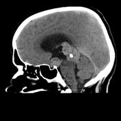
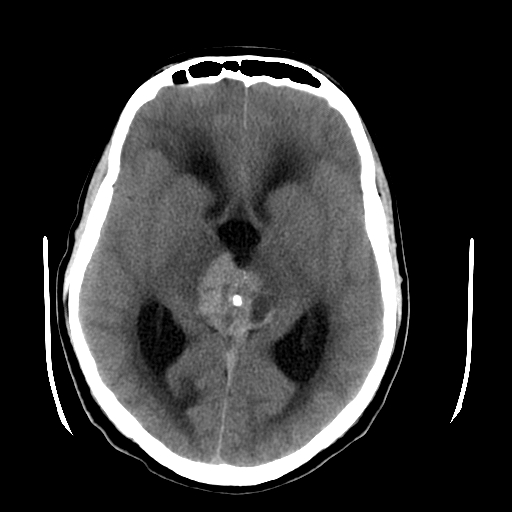
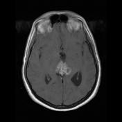
A large lobulated mass is centered on the pineal gland, engulfing the pineal calcifiation. It is somewhat hyperdense compared to adjacent brain. A further smaller mass is seen in the floor of the third ventricle. The midbrain is distorted, compressed and demonstrates low density suggestive of edema. Obstructive hydrocephalus is present.

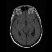
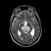
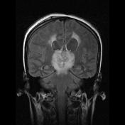
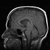
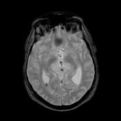
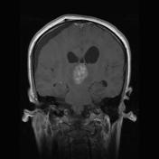
An EVD has been placed and the size of the ventricles has reduced. A subdural collection has developed, which is blood stained. The masses seen on CT are more easily seen on MRI. They are heterogeneous with prominent variegated enhancement, and a moderate amount of edema extending into the adjacent parenchyma, particularly the brain stem.
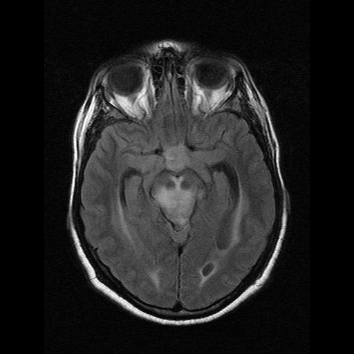
On gradient echo imaging the pineal calcification is confirmed to be placed centrally within the mass (engulfed).
This patient went on tho have a craniotomy and a biopsy of the pineal region mass confirming the diagnosis of a germinoma.
Case Discussion
This case illustrates fairly typical appearances of a germinoma with involvement of both the pineal region and the floor of the third ventricle / suprasellar region.


 Unable to process the form. Check for errors and try again.