
Presentation
Acute memory loss for past 3-4 days, confused by simple tasks.
Patient Data
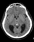

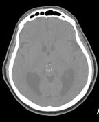
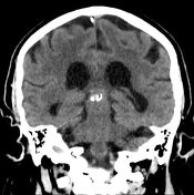
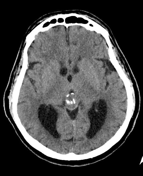
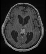
No evidence of intracranial bleed or brain infarct.
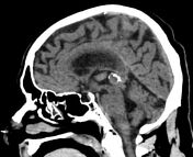
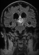
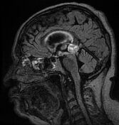
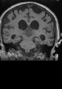
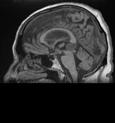
Heterogeneous mass lesion at the location of the pineal gland, measuring 15 x 20 x 13 mm (TR x AP x CC), with internal calcification and peripheral calcification at its anterior edge.
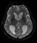
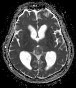
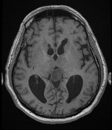
The lateral ventricles are dilated. The third ventricle is narrow, despite apparent Sylvian aqueduct compression.
The pituitary gland is flattened across the dorsum sellae.

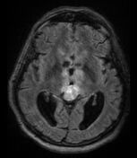
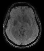
Movement-related artifacts are present in many sequences.
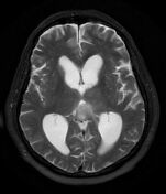
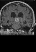
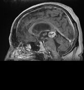
Pineal mass lesion measuring 2 cm in largest diameter, with a lobulated border. The mass is slightly heterogeneous, mostly intermediate-low signal intensity on T2WI and T1WI. It extends anteriorly in the paramedian thalami on both sides of the third ventricle and into the mammillary bodies and midbrain. The lesion enhances slightly heterogeneously after Gadolinium injection. Foci of hemorrhage in the pineal mass and area of restricted diffusion in the lateral aspect of the lesion. The pineal mass compresses the third ventricle, resulting in moderate dilation of the lateral ventricles - obstructive hydrocephalus. High FLAIR signal around the lateral ventricles, denoting interstitial cerebral edema.
The differential diagnosis includes pineal parenchymal tumor of intermediate differentiation (PPTID) and glioma (less likely). Low probability of germinoma and papillary tumor in light of the patient's age.
Case Discussion
A biopsy was obtained from the periventricular part of the mass. Histopathology report follows:
High grade glioma compatible with glioblastoma, IDH-wildtype, WHO grade 4.
A small fragment from a neoplasm composed in part from small mononuclear cells and in part from large cells with extensive eosinophilic cytoplasm and cytological atypia. Mitotic activity seen. Necrotic foci seen. Inconclusive microvascular proliferation. The cells are not arranged in any particular order. Scattered stromal calcifications. Immunohistochemical staining: positive for olig2, GFAP; negative for IDH1, EGFR, H3K27M, NF, SYN, and KERMNF116. Staining for ATRX was negative (loss of expression). Very few scattered cells stained positive for p53. ~10% of cells were positive for Ki67.
A ventriculoperitoneal shunt (VPS) was inserted into the left ventricle.
Pineal region glioblastoma is exceedingly rare.


 Unable to process the form. Check for errors and try again.