
Presentation
Headaches.
Patient Data


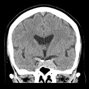
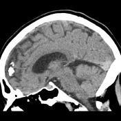
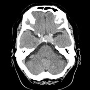
A soft tissue density mass on the left side of the sella is associated with bony remodelling and on post-contrast imaging appears to enhance similarly to the adjacent internal carotid artery.

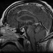
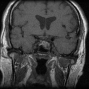
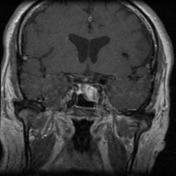
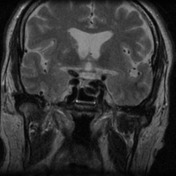
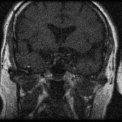
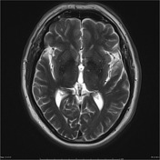
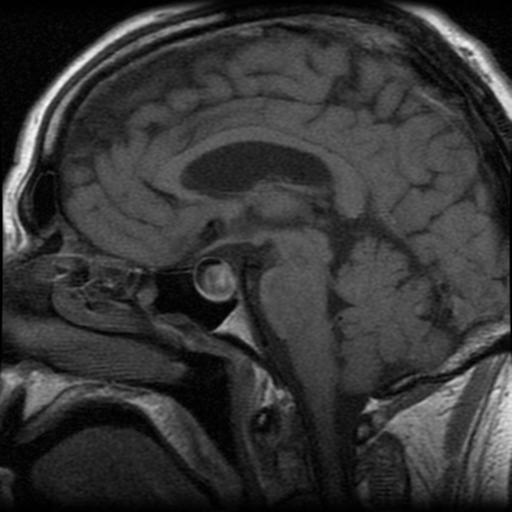
There is an ovoid mass within the sella and left cavernous sinus. The mass is closely related to the left cavernous internal carotid artery but does not appear to be causing arterial stenosis. Within the mass there are prominent flow voids. No solid component. The pituitary and infundibulum are displaced towards the right. No diffusion restriction seen.
Conclusion: Left sellar/suprasellar mass almost certainly represents an ICA aneurysm arising from the left cavernous ICA.


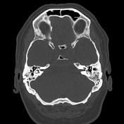
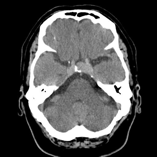
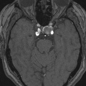
CTA demonstrates a large medially projecting aneurysm.
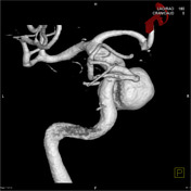
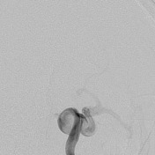

A very large left cavernous ICA aneurysm measuring approximately 17.5 x 15 mm with a narrow neck estimated 6 mm projecting inferomedially. Fetal PCOM anatomy.
Case Discussion
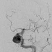
The aneurysm went on to be coiled.
It is essential to remember to ask yourself when looking at a pituitary region mass, particularly on MRI where clot and flow-related artifacts can be confusing, "Could this be an aneurysm?", as not doing so and only finding out once a surgeon has attempted to resect the 'adenoma' can be disastrous.


 Unable to process the form. Check for errors and try again.