
Presentation
Chronic headache, mild vision loss, galactorrhea, and fatigue.
Patient Data
Age: 55 years
Gender: Female
From the case:
Pituitary macroadenoma

Download
Info

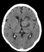
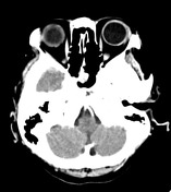
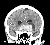
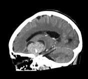
CT images show a large mostly solid mass with some cystic areas is seen arising from the sellar fossa that looks expanded. The mass is seen extending to the suprasellar cistern, compressing the base of the frontal lobe, hypothalamic region, optic chiasm, and abutting the floor of the third ventricle. The lesion shows moderate enhancement and encases both carotid arteries and the left middle cerebral artery.
Case Discussion
Imaging findings suggestive of pituitary macroadenoma. Differential diagnosis includes; pituitary carcinoma, metastasis, or papillary craniopharyngioma. Sometimes they can be indistinguishable on imaging.


 Unable to process the form. Check for errors and try again.