
Presentation
The patient is asymptomatic at the time of MRI. She has a known history of primary amenorrhoea and panhypopituitarism.
Patient Data


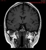
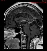
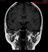
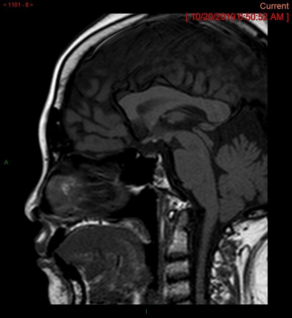
The sella is predominantly CSF filled with a small and flattened anterior pituitary gland along the sellar floor with a height of 0.2 cm. On sagittal T1 thin sequence, no normal posterior pituitary bright spot is seen in the pituitary fossa. There is a note of a punctate T1 hyperintense focus at the level of median eminence likely relating to an ectopic posterior pituitary. The pituitary stalk is not identified.
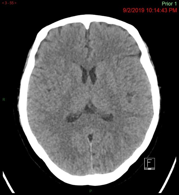
No CT evidence of acute intracranial haemorrhage, large acute territorial infarct, focal mass lesion or oedema.
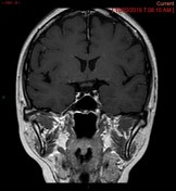
- fluid-filled sella
- poorly developed and sclerotic bilateral mastoid air cells
- high riding right jugular bulb with dehiscence
- lateralised bilateral sigmoid sinuses, more pronounced on the right side
Case Discussion
Patient's Lab Test results:
- prolactin 7.40 ng/mL (Reference range = Not-pregnant: 2.8 - 29.2 ;Pregnant: 9.7 - 208.5; Post Menopausal: 1.8 - 20.3)
- oestradiol 19.35 pg/mL (Reference range = Follicular phase: 19.5 - 144.2; Ovulation phase: 63.9 - 356.7; Luteal phase: 55.8 - 214.2; Post-menopausal: <32.2)
- luteinizing Hormone (LH) < 0.07 mIU/mL (Reference range = Follicular phase: 1.9 - 12.5; Ovulation phase: 8.7 - 76.3; Luteal phase: 0.5 - 16.9; Post-menopausal: 15.9 - 54.0; Pregnant : Less than 1.5; Contraceptives : 0.7 - 5.6)
- follicle Stimulating Hormone (FSH) 0.95 mIU/mL ( Reference range = Follicular phase: 2.5 - 10.2; Ovulation phase:3.4 - 33.4)
Case Discussion
Findings of hypoplastic anterior pituitary gland, ectopic posterior pituitary and unidentifiable/absent pituitary stalk are imaging findings suggestive of pituitary stalk interruption syndrome.


 Unable to process the form. Check for errors and try again.