
Presentation
Been out with friends, slumped unresponsive drowsy, slow response to r/o acute intracranial pathology.
Patient Data

Suspicion of macroadenoma of the pituitary gland - differential diagnosis of a cerebral artery aneurysm.
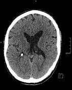
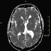
Cavum septi pellucidi et vergae.
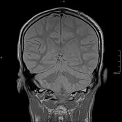
The ventricles, sulci and basal cisterns are within normal limits. No skull fracture.
Pansinusitis. Aerated mastoid cells.
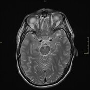
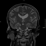
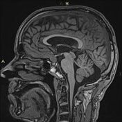
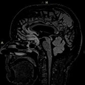
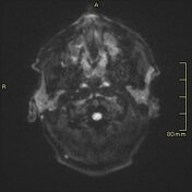
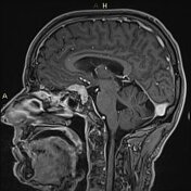
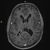

Homogeneously, avidly enhancing lesion with a dural tail arising from the planum sphenoidale. The posterior aspect abuts the superior pituitary gland and infundibulum. The lesion abuts both intracavernous ICAs and the optic chiasm.
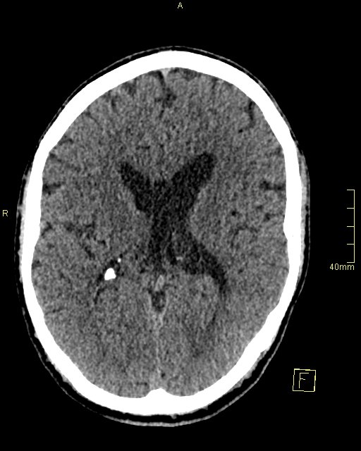
Cavum septum and cavum vergae.
Mucosal thickening in the maxillary and ethmoidal sinuses.
Case Discussion
A planum sphenoidale meningioma - I always think it has a rather 'slug-like' appearance as it drapes itself over the planum sphenoidale.
Radiologists seem to love radiologically inspired animal descriptors although the 'slug sign' is perhaps one that won't get any traction!


 Unable to process the form. Check for errors and try again.