
Presentation
Chronic sinusitis, history of nasal and sinus surgery (details unavailable)
Patient Data
Age: 40 years
Gender: Male


Download
Info

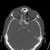
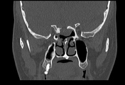
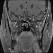
- central nasal septal defect
- partial right middle turbinectomy
- polypoidal mucosal thickening seen in right maxillary sinus ostium along with circumferential sinus mucosal thickening
- air fluid levels seen in bilateral ethmoid air cells along with mild mucosal thickening in right frontonasal drainage
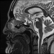
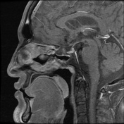
- bony defect of the right posterior planum sphenoidale with well defined sac and convex inferior margins projecting into the sinus cavity
Download
Info

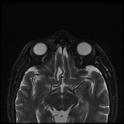
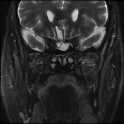
- posterior right gyrus rectus encephalomalacia with herniation of part of gyrus through the defect with an intact meningeal lined sac of CSF protruding into the sinus cavity.
Case Discussion
Planum sphenoidale defect with sinus communication of neuroparenchyma can occur in sinus surgeries. In particular, they may occur during middle turbinectomy and uncinectomy depending on the anatomic configuration of the latter. This may result in CSF rhinorrhoea, and should be brought to the attention of the ENT surgeon, for urgent planum sphenoidale repair, as life threatening cranial spread of infection can occur.


 Unable to process the form. Check for errors and try again.