
Presentation
Dorsal deformity, scoliosis and walking difficulty, without neurological commitment, has no personal pathological history.
Patient Data
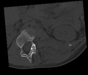

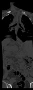
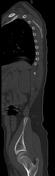
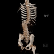
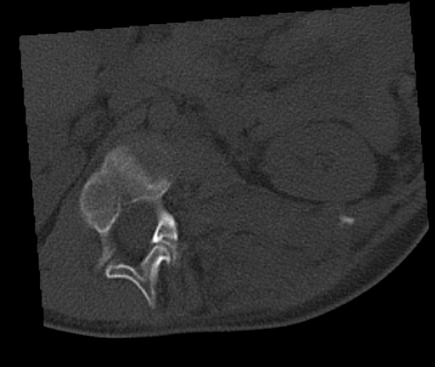
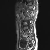
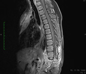
Severe thoracolumbar scoliosis due to a well-defined, lobulated left paravertebral mass, isodense to adjacent soft tissues.
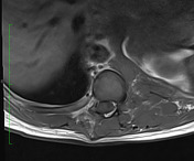
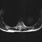
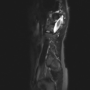
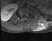
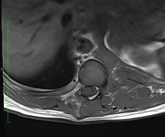
Mixed composition, heterogeneous on all sequences, with cystic areas that conflate giving it the appearance of a "bag of worms".
Origin at the right vertebral conjunction orifice level T11-T12, covering from T10 to L1 level. It extends through the fascia of the erector muscle of the back and quadratus lumborum, conditioning the volume effect that displaces psoas muscle, posterior para-renal space and crura, in addition, associated bone remodelling is observed,
Case Discussion
This case shows classic findings of a mass-like predominantly hyperintense with some whorled hypointensity centrally in an extradural paraspinal localisation with involvement of spinal root that emerges at and widens the T11-T12 neural exit foramen.
The images are particularly representative and lead to the diagnosis of neurofibromatosis type I. The mass was biopsied, confirming plexiform neurofibroma on histopathology.


 Unable to process the form. Check for errors and try again.