
Presentation
Bilateral flank pain, nausea and vomiting, and hematuria.
Patient Data
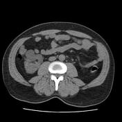

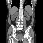
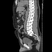

There are numerous cysts in both kidneys with hyperdense foci within some of the cysts compatible with hemorrhage. There are also a few scattered small calculi within the kidneys and a few small hypodense foci in hepatic lobes more consistent with small liver cysts.
Case Discussion
A case is a 50-year-old man who came to the emergency room with the clinical symptoms of flank pain and hematuria. The ureteral stones suspected and non-contrast abdomen and pelvic MDCT were requested. On performed MDCT, numerous cysts of different sizes and a few scattered small stones with small hyperdense foci compatible with hemorrhage within some of the cysts in both kidneys were detected. A few small hypodense foci in hepatic lobes compatible with small cysts are also seen. Decreased kidneys' parenchyma interposed the cysts were also seen. Findings were compatible with polycystic kidney disease.
A high-resolution ultrasound exam, MDCT, and MRI all can be useful for the diagnosis of polycystic kidney disease patients but the MRI is considered highly sensitive and specific in some of the recent studies for diagnosis and also a follow-up of kidneys status autosomal dominant polycystic kidney disease. These patients are also at greater risk of intracranial arterial aneurysm and MRA also is the preferred imaging modality for follow-up of the patients from this point of view.


 Unable to process the form. Check for errors and try again.