
Presentation
Headache. Previous MRI of the brain revealed pontine central abnormal signal with radiological possibilities of MS or pontine glioma. MRS was requested for further assessment.
Patient Data


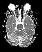
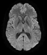
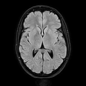
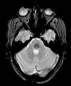
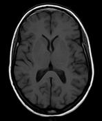
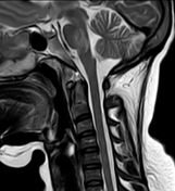
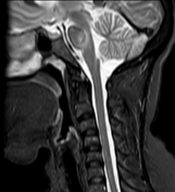
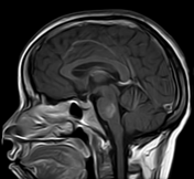
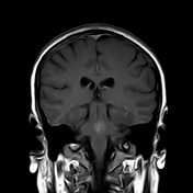
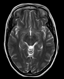
The pons show a central patchy area of abnormal increased signal on T2 WI. It elicits a subtle low signal on T1 and a high signal on FLAIR WI. It's hypointense on DWI. Corresponding blooming artifact on GRE WI. The lesion shows patchy post-contrast enhancement with a brush-like appearance on the post-contrast series. No surrounding edema nor a mass effect. No significant elevation of the choline peak on MRS.
Central linear draining vein/DVA is seen within the posterior central aspect of the lesion which elicits a high signal on GRE, low signal on T2 and STIR, and linear contrast enhancement.
Opinion: radiological features with the characteristic blooming on GRE and patchy contrast enhancement are impressive in pontine capillary telangiectasia.
Case Discussion
CNS capillary telangiectasias are small, asymptomatic low-flow vascular lesions of the brain. They are considered the second most common vascular anomaly after developmental venous anomalies on imaging.
Typical radiological features include subtle to slightly high signal on T2 and FLAIR WI, blooming artifact on GRE, and patchy contrast enhancement 1. This hypointense signal on GRE is likely secondary to the slow blood flow within the lesion, which causes an increased deposition of desoxyhemoglobin within the lesion resulting in a signal loss in T2* images 3. Characteristic hypointense signal on DWI has also been reported 3.
Capillary telangiectasias are almost always asymptomatic. However, in some cases, it might be associated with DVAs. McCormick et al. suggested that elevated venous pressure in a DVA leads to dilated microvasculature, representing acquired telangiectasia that evolves toward a cavernous malformation 2. Given this hypothesis, the symptoms are presumed to be primarily caused by the outflow restriction of the DVA, not by the secondary capillary telangiectasia 2. Abla et al. 4 concluded, that DVAs, telangiectasias, and cavernomas represent a spectrum within a single pathological entity, and considered that cavenomas are a more developmentally mature form of capillary telangiectasias, albeit the product of the same pathologic process 4.


 Unable to process the form. Check for errors and try again.