
Presentation
The patient is a known case of autoimmune hepatitis. She had a splenic artery aneurysm that had been embolized by an interventional radiologist.
Patient Data
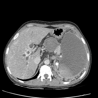
Evidence of chronic portal vein thrombosis with numerous small venous collaterals at porta-hepatis (cavernous transformation of portal vein), around the bile duct confluence, central bile ducts, and common hepatic duct are evident.
Mild intrahepatic biliary dilation is present down to the hilum. The porta-hepatis collaterals mimic an enhancing hilar mass such as klatskin tumor. Chronic intrahepatic and extrahepatic portal thrombosis, equivalent enhancement to the vessels, and no bile duct luminal obliteration favor portal cholangiopathy.
Evidence of coil embolization and thrombosis of the proximal splenic artery aneurysm and the entire splenic artery course is visible. The enlarged spleen shows evidence of infarction because of the splenic artery embolization/thrombosis. Multiple short gastric and peri-splenic collaterals due to portal hypertension are visible too.
Case Discussion
The patient underwent follow up imaging. Laboratory data and imaging features were stable on follow up.


 Unable to process the form. Check for errors and try again.