
Presentation
Hypoechoic mass lesion in Right lobe of liver on USG.
Patient Data
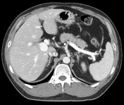

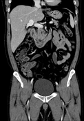

About 1.8 cm sized saccular aneurysmal dilatation of the right main portal vein. No evidence of liver cirrhosis, portal vein anomaly, or portal vein thrombosis. Incidental finding of haemangiomas in the left lobe of the liver (4 cm, 1.8 cm).
Case Discussion
Portal venous aneurysms (PVAs) are rare venous aneurysms. The mean diameter of a healthy portal vein varies considerably, with a maximum diameter of 15 mm in healthy subjects. They are more often as an incident imaging finding but sometimes they could come with abdominal pain or more often as an incident imaging finding. The aetiology and the mechanism of formation of PVA remain ill-defined. PVA may be associated with various complications: thrombosis, aneurysmal rupture, inferior vena cava obstruction, or duodenal compression. A conservative treatment showed a satisfying clinical and radiological response, however, surgical and endovascular options can be considered.
In this case, the right main PV saccular aneurysmal dilatation was found at first on ultrasonography with haemangiomas in the left lobe of the liver. After that, we confirmed portal vein aneurysm and haemangiomas on CT. The patient was asymptomatic and had not experienced any complications.


 Unable to process the form. Check for errors and try again.