
Presentation
Abdominal pain and vomiting
Patient Data


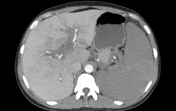
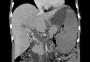
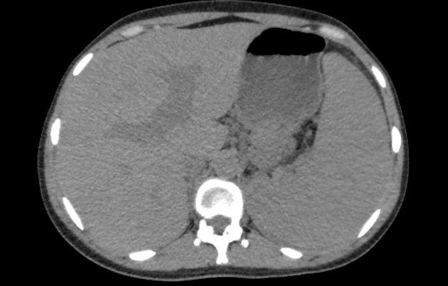
The liver is mildly enlarged, with multiple geographic mainly right lobe hypodense non-enhancing patches (perfusion difference).
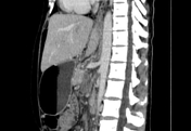
The portal vein (with its right and left main branches) and splenic vein are distended with luminal thrombosis with mild enhancement of portal venous wall.
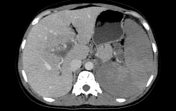
The superior mesenteric and inferior mesenteric veins, as well as their branches, are completely thrombosed (mesenteric venous occlusion).
The spleen is markedly enlarged (measuring 22 cm in bipolar diameter - coronal plane) showing multiple wedge-shaped non-enhanced areas representing splenic infarcts.
Case Discussion
The portal venous system consists of the mesenteric, splenic and portal veins. Occlusion of portal veins causes congestion of liver, spleen and bowel loops due to increased venous pressure and the development of venous collaterals.
Acute thrombosis may be difficult to detect in non-contrast basis as the thrombus may be iso or hypodense and not usually be hyperdense. The diagnosis is made on the portal venous phase of contrast-enhanced studies.


 Unable to process the form. Check for errors and try again.