
Presentation
Elite athlete. Ongoing shoulder pain 2 weeks post-injury despite treatment.
Patient Data


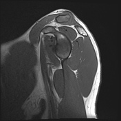
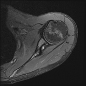
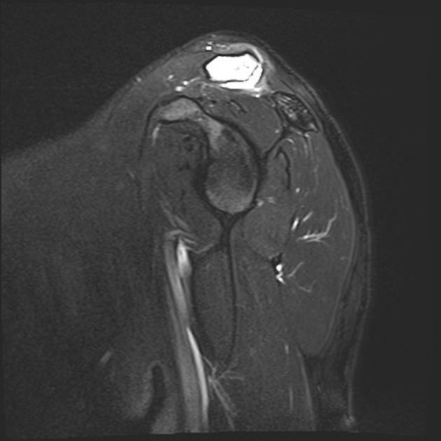
Elevated distal clavicle but not beyond the superior acromion; no anteroposterior displacement. Acromioclavicular effusion with capsular oedema. Marked oedema on the distal clavicle with irregularity of distal articular surface and loss of normal low T1 signal. Mild oedema within the adjacent acromion. Coracoclavicular ligament is intact.
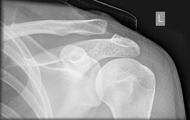
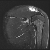
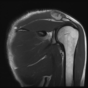
Similar alignment to the prior with mild elevation of the distal clavicle compared to the acromion. No articular erosion.
Case Discussion
Distal clavicular osteolysis has a myriad of causes with trauma being one of them. While the acromioclavicular joint injury is a Rockwood type I/II, which typically heal well with conservative measures, in this case the clinical course has become complicated.


 Unable to process the form. Check for errors and try again.