
Presentation
Acute onset of vertigo, nausea and vomiting.
Patient Data
Age: 35 years
Gender: Male
From the case:
Posterior inferior cerebellar artery (PICA) infarct


Download
Info

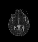
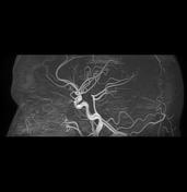
There is a large left cerebellar infarct involving the posterior inferior cerebellar artery (PICA) territory seen as a large area of low signal on T1, high signal on T2 and FLAIR with restricted diffusion. A mass effect is noted on the 4th ventricle with moderate dilatation of the 3rd and lateral ventricles. On MRA 3D-TOF the right PICA is well-visualized arising from the vertebral artery while the left is not visualized.
Case Discussion
MRI appearances in keeping with an acute PICA territory infarct.


 Unable to process the form. Check for errors and try again.