
Presentation
Bloody diarrhea and obstructive jaundice.
Patient Data
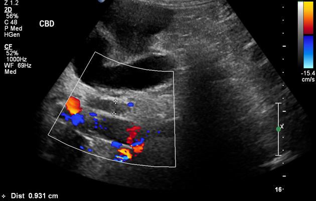
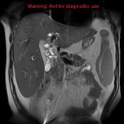
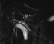
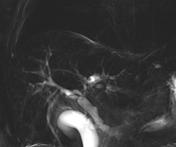
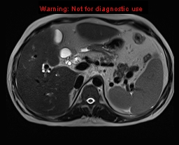
CBD is dilated, around 9 mm in maximum diameter.
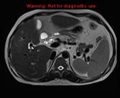
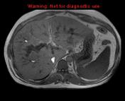
Hepatosplenomegaly is noted. Fine coarse echotexture of the liver and border irregularity reflecting mild cirrhosis. Multiple porta hepatis lymph nodes.


The liver is enlarged and demonstrates fine surface nodularity suggestive of early cirrhosis.
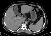
CBD is dilated, around 9 mm in maximum diameter and demonstrates mural thickening and enhancement. No mass lesion is seen at the distal CBD or head of the pancreas. Note is made of moderate dilatation of the left intrahepatic biliary radicles with focal cystic dilatation of left hepatic duct. There is mild dilatation of the right intrahepatic radicles centrally. No definite mass lesion could be identified. Couple of subcentimeter porta hepatis lymph nodes is seen. The pancreatic duct is not dilated.
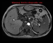
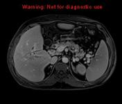
The spleen is enlarged, measuring 18 cm in the CC dimension.
Mucosal thickening of the right colon involving the cecum, ascending and hepatic flexure. Mild mucosal thickening is also seen in the terminal ileum. A couple of subcentimeter mesenteric lymph nodes are seen as well. No mesenteric fat strandings are seen. No free fluid is seen. Findings are highly suggestive of burn out or chronic colitis.

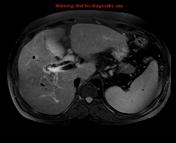
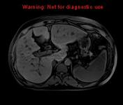

The liver is enlarged and demonstrates fine nodularity of the surface and prominent fissures representing cirrhosis. No focal hepatic lesion otherwise is demonstrated.
The gallbladder appears unremarkable. No gallstone seen.
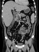
The CBD is dilated measuring 9 mm in the maximum diameter. CBD wall thickening and enhancement are seen. An area of cut off is noted at the distal CBD representing tight stricture.
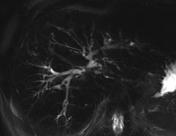
Intrahepatic biliary duct dilatation is noted, more pronounced at the left hepatic lobe, which is disproportionate in regard to dilatation. Another area of focal tight stenosis is noted at the intrahepatic bile duct at the left hepatic duct near the confluence with the common hepatic duct. The intrahepatic biliary ducts are abnormal and demonstrate multifocal areas of stenosis and dilatation giving the beaded appearance.
Splenomegaly. Multiple enlarged lymph nodes are noted at the porta hepatis, aortocaval, peripancreatic and gastrohepatic region.
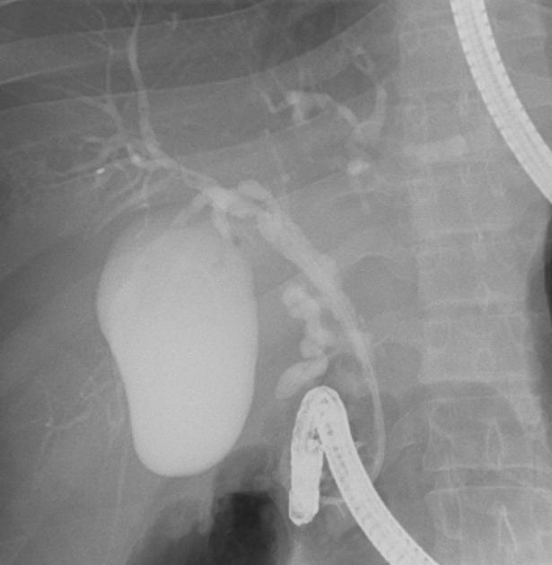
Dilated CBD and intrahepatic biliary radicals with focal dilatations and strictures.


 Unable to process the form. Check for errors and try again.