
Presentation
Abdominal pain and weight loss.
Patient Data
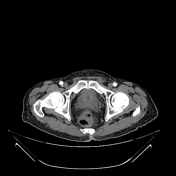
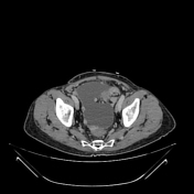
CT scan showed omental masses and the biopsy revealed C- kit positive high-grade extra-intestinal GIST. The baseline and follow-up scans six months later are presented to assess the response to imatinib therapy.

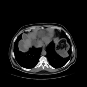

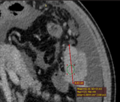
The first target lesion was a confluent omental mass measuring 48mm (single largest diameter), of 82 HU mean density
The second target lesion was an omental mass measuring 10 mm (single largest diameter), of 97 HU mean density.
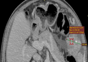
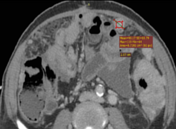

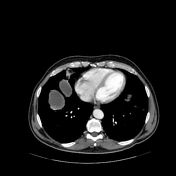
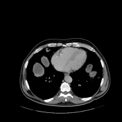
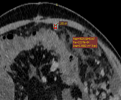
The first target lesion now measures 58.2 mm (single largest diameter), of 86 HU mean density
The second target lesion now measures 22.7 mm (single largest diameter), of 90 HU mean density.
Nontarget lesions:
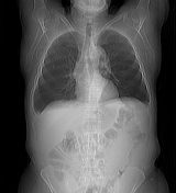
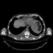
Bilateral moderate pleural effusion more on the right side (new)
Enhancing peritoneal thickening (unequivocal progression)
Moderate ascitic fluid is seen in the pelvis, right paracolic gutter, and right subphrenic space, the root of the jejunal mesentery (less than the previous scan), no hemoperitoneum
-
Innumerable, mostly spherical and deforming, vascular peritoneal and omental implants, some are necrotic confluent and are heterogeneously enhancing, multiple serosal implants thickening loops of bowel, no obstruction… peritoneal sarcomatosis(unequivocal progression)
Case Discussion
The sum of baseline scan target lesion diameters is 58mm with a mean density of 89.5 HU, and the sum of current target lesion diameters is 80.9mm with a mean density of 88.0 HU thus the change in tumor size is 39.5 % with -1.7 % change of tumor density indicating progressive extra-intestinal GIST according to Choi Response Criteria
Primary GISTs could affect the peritoneum involving the mesentery or omentum. It appears heterogeneous due to internal hemorrhage, necrosis, and cystic change; the solid component enhances upon contrast administration. It may be indistinguishable from malignant fibrous histiocytoma, leiomyosarcoma, liposarcoma, and other sarcomas including metastasizing GISTs from the gastrointestinal tract on cross-sectional imaging 1.


 Unable to process the form. Check for errors and try again.