
Presentation
Pain in the left arm.
Patient Data
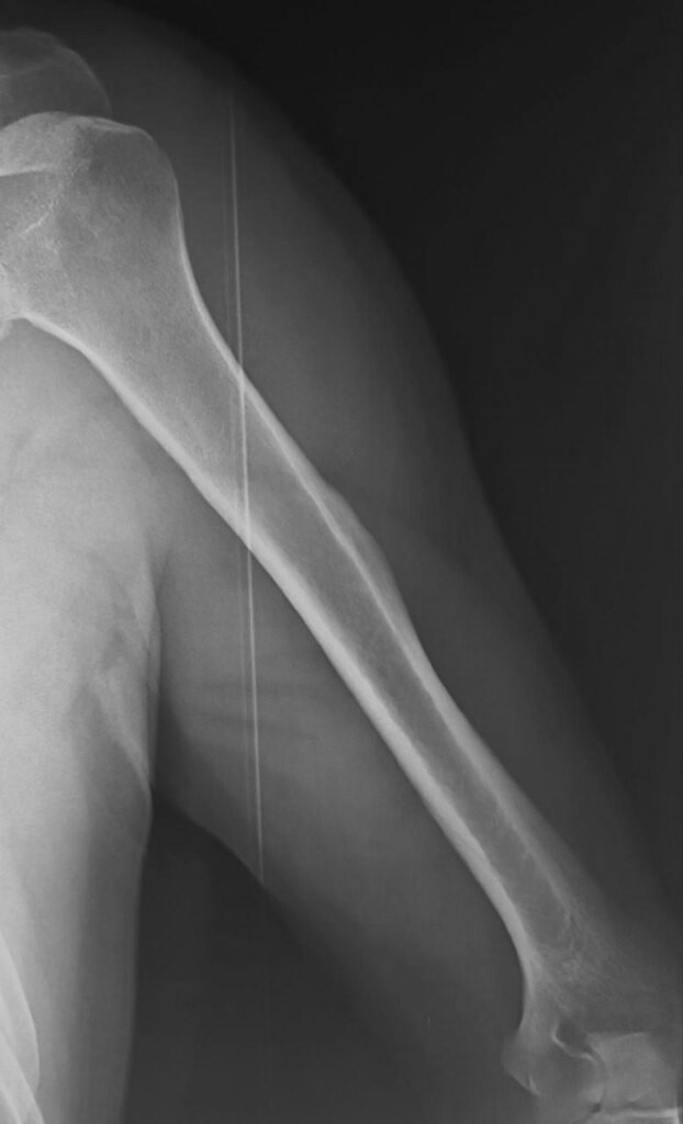
Cortical thickening in the proximal humerus at the deltoid muscle insertion site with internal lucency.


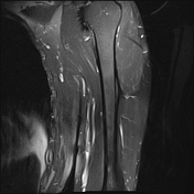
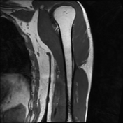
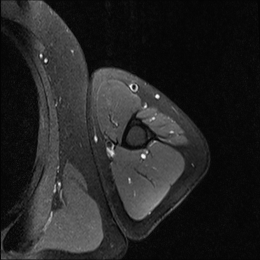
Evidence of cortical thickening in the proximal humerus at the deltoid insertion site with central bone marrow signal intensity, no associated bone marrow oedema or soft tissue lesion.
Case Discussion
The imaging features are highly suggestive of pseudotumour deltoideus (prominent deltoid muscle insertion in the proximal humerus) which is a rare benign anatomical variant that should not be mistaken for tumour or infection. Cortical thickening with central radiolucency and moderate T2 signal intensity is typical and biopsy should be avoided.
Patients may present with discomfort, pain, or without any symptoms 2.
This term was initially introduced in 2001 by Morgan et al 1.


 Unable to process the form. Check for errors and try again.