
Presentation
General fatigue, lower chest and upper abdominal pain, and dyspnea.
Patient Data


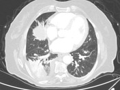
Multiple contrast media filling defects in the left upper and lower main lobar arteries, and in multiple right and left segmental branches.
Dilated right and left main pulmonary arteries.
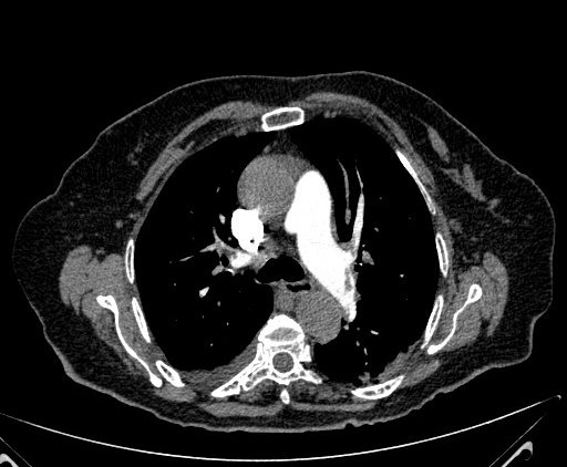
Peripherally consolidative areas in both lungs with their supplying arteries are showing contrast filling defects, along with being:
- the consolidation in the right lower lobe showing variable postcontrast attenuation, with internal air lucencies (bubbly consolidation and suspected cavitation)
- the consolidation in the superior segment of the left lower lobe.is wedge-shaped
The heart appears enlarged.
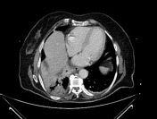
The visualized aorta appears unremarkable apart from calcified atheromatous plaques.
Dilated intrahepatic biliary tree, and a lobulated cystic lesion in right hepatic lobe, are noted.
Case Discussion
Multiple bilateral pulmonary emboli and infarcted areas of lung.
In this case, the consolidations are favored to be considered infarctions by:
- consolidative-supplying artery thromboembolism (in both lungs)
- postcontrast hypoattenuating lung parenchyma with small air foci (bubble consolidation) and suspected cavitation (in the right lung)
- peripherally wedge-shaped consolidation (in the left lung)
The cystic lesion in the liver could be a simple cyst or biloma, and the intrahepatic biliary dilatation should be further evaluated as well as searching for the cause of the pulmonary emboli.


 Unable to process the form. Check for errors and try again.