
Presentation
Premature newborn (born at 25 weeks) on high frequency jet ventilation.
Patient Data
Series of chest and abdominal radiographs:
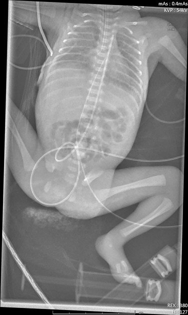
First one obtained after ET tube placement, central lines and administration of a dose of surfactant.
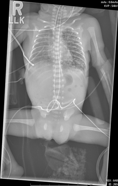
Tubes and lines:
Tip of the ET tube in right main bronchus, deep position of the umbilical arterial catheter (at T2) level, deep position of the umbilical venous catheter (high right atrium).
Lungs are mildly hypoinflated. Bilateral diffuse hazy opacities in the lungs. Absence of typical surfactant deficiency findings (low volume lungs and granular appearance of the lungs) is due to surfactant administration before taken the image.
The others were taken at 10, 20, 24 and 36 hours after birth.
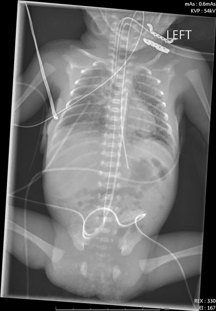
10 hrs image:
Tubes and lines: placing an NG tube in the interim. ET, UAC , UVC are adjusted.
Lungs are more expanded than prior image. Multiple patchy opacities scattered throughout left lung and right perihilar region with non-branching linear radiolucencies more prominent in the left lung.
Findings are consistent with early development of pulmonary interstitial emphysema
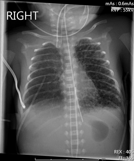
Tubes and lines are in satisfactory positions.
Numerous diffuse bubbly and linear radiolucencies occupying most of the left lung field. Similar radiolucencies, but less extensive, also seen in the right perihilar region and right upper lobe.
Findings are consistent with pulmonary interstitial emphysema.
Similar findings to before:
Findings are consistent with pulmonary interstitial emphysema in the left lung, right perihilar region and upper lobe.
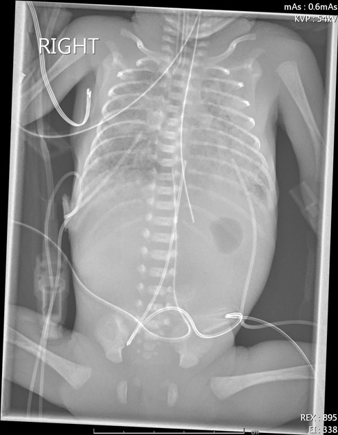
Tubes and lines are in a satisfactory positions.
Significant improvement in the bilateral pulmonary interstitial emphysema.
There is interval worsening of the diffuse confluent opacities.
Case Discussion
This is a typical case of pulmonary interstitial emphysema (PIE) in a premature newborn who is on mechanical ventilation to treat surfactant deficiency. It developed in the first day of life and significantly improved by the 36 hrs image.


 Unable to process the form. Check for errors and try again.