
Presentation
History of seizures.
Patient Data
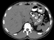

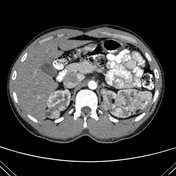
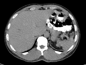
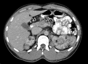
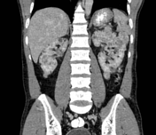
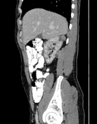

Multiple lesions of varying sizes with mixed fat density and soft tissue component are seen in both kidneys. No evidence of perinephric hematoma.
Case Discussion
This is a typical case of angiomyolipomas.
The patient presented with a history of seizures from childhood, accompanied by intellectual disability and facial angiofibromas. A CT brain scan revealed multiple bilateral subependymal calcific foci, consistent with subependymal hamartomas of varying sizes. A CT abdomen was subsequently performed, showing bilateral angiomyolipomas, although the patient did not report any abdominal symptoms. Despite attending multiple dermatology and neurology clinics since childhood, a final diagnosis of tuberous sclerosis was only made at the age of 40.
While the majority of angiomyolipomas (approximately 80 percent) are sporadic, the remaining 20% are associated with phakomatoses, most notably tuberous sclerosis 1. This case underscores the importance of a multidisciplinary approach to diagnosis and management, as it can lead to a timely and accurate diagnosis, potentially preventing delayed recognition and treatment.


 Unable to process the form. Check for errors and try again.