
Presentation
Left flank pain and microscopic haematuria for six months.
Patient Data


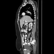
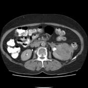
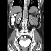
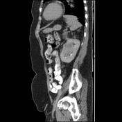

Large solid-necrotic hypervascular and lobulated margin mass with heterogeneous enhancement and some cystic foci and axial width up to 80 x 75 mm and height up to 80 mm in middle to the lower pole of the left kidney extended within perirenal space and with invasion to related pyelocaliceal system and renal sinus fat is seen. There is not any obvious extension within the renal vein and IVC and no obvious paraaortic lymph node. No obvious solid organ metastasis is seen.
Case Discussion
The case illustrates contrast-enhanced MDCT features of pathology-proved non-metastatic clear cell renal cell carcinoma. Clinical management is usually based on renal cell carcinoma TNM staging. Flank pain and haematuria are usually seen in large tumours in particular cases 1.


 Unable to process the form. Check for errors and try again.