
Presentation
For investigation of tingling in her face. Pre-op cataract surgery.
Patient Data
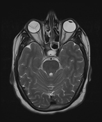

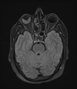

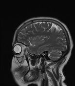

The right globe shows a linear low signal membrane in the vitreous chamber extending anteriorly in a V-shape with anterior insertion into the ora serrata and posterior convergence at the optic disc. Sub-retinal intermediate T1 signal denotes hemorrhage.
Right intra-ocular lens implant.
Case Discussion
The diagnosis of retinal detachment was made clinically and confirmed on the brain MRI. The patient underwent cataract surgery but the retinal detachment was not treated.
Retinal detachment is limited anteriorly by the ora serrata. In contrast, a choroidal detachment extends beyond the ora serrata. Posteriorly, a retinal detachment converges on the optic disc, while a choroidal detachment diverges at the disc.


 Unable to process the form. Check for errors and try again.