
Presentation
Right lower pleuritic chest pain
Patient Data


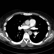
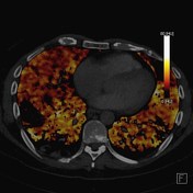
The lateral basal segmental branch of the right lower lobe artery demonstrates a filling defect with, with the corresponding iodine map confirming. Consistent with a pulmonary embolism.
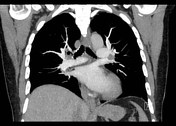
Suboptimal enhancement of the pulmonary trunk with an HU of 178. The monochromatic data set has increased the enhancement of the pulmonary trunk with an HU of > 200.
Case Discussion
The following case is a great example demonstrating the benefits of dual energy CT, in an earlier time scans that produce lower Hounsfied units over the pulmonary artery would require repeating until the scan was satisfactory.
Although this scan is borderline adequate, utilising the monochromatic data set of 50 KeV has better demonstrated the contrast filled structures. The monochromatic energy is closer to the K-edge of iodine, and therefore iodine-enhanced structures will display better attenuation values.
Not every failed CTPA can be 'saved' using this method, yet it should be considered before re-scanning 1,2.


 Unable to process the form. Check for errors and try again.