
Presentation
Collapse with left sided weakness, neglect and poor speech. Imaged between 60-90 minutes from onset. NIHSS 21.
Patient Data
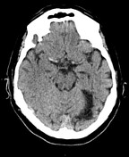

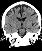
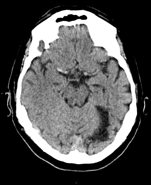
Hyperdensity in right M1 vessel. Reduced attenuation of right insular cortex and lentiform nucleus. No other established MCA territory infarct. ASPECTS 8.
Old left PCA territory / occipital infarct.
No haemorrhage or mass.
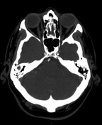
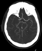
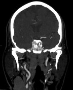
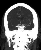
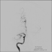
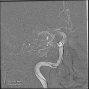
Angiogram confirms right M1 occlusion. Tortuous cervical ICAs with minor calcific but non-flow limiting stenosis of the carotid bulbs.
-
IV tPA administered on transfer to neurointervention suite.

Onset-to-skin puncture time: 136 minutes
Onset-to-reperfusion time: 157 minutes
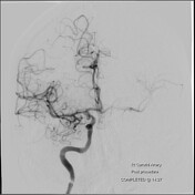
Right M1 clot retrieved on first pass; TICI 3.
NIHSS 3 immediately post-treatment.
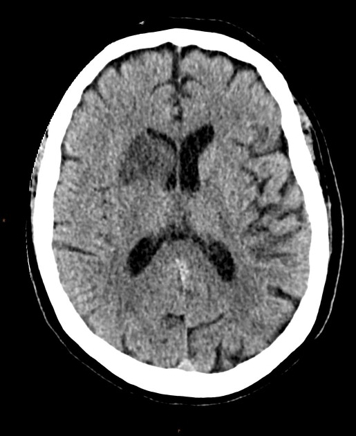
Maturing infarct in right lenticulostriate territory, including caudate head, anterior limb of internal capsule, lentiform nucleus. Central speckled hyperattenuation considered contrast staining. No haemorrhagic transformation.
Case Discussion
This patient presented with a high NIHSS, some lateralising signs and general confusion with speech disturbance and neglect.
Unenhanced CT and CTA were performed (the patient was considered too agitated for multiphase CTA / perfusion). A right M1 occlusion was confirmed, with early insular / lenticuostriate infarct.
The clot was evacuated on first pass, with good clinical and radiographic outcomes.
-
Case courtesy of Dr Alberto Nania, INR consultant


 Unable to process the form. Check for errors and try again.