
Presentation
Cyclical pain and vomiting
Patient Data


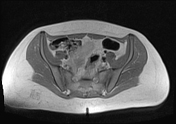
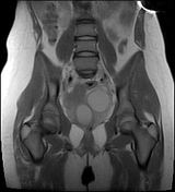
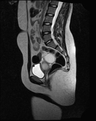
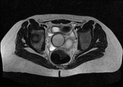
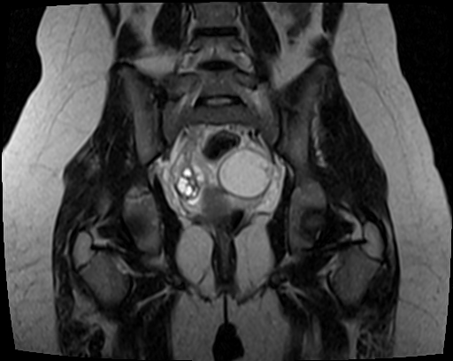
Normal-shaped external uterine fundus. On coronal T2 images, a hypointense uterine septum is seen longitudinally and asymmetrically dividing the uterus into two non-communicating hemicavities, a small right cavity, and a larger left cavity. The left endometrial cavity is distended with blood seen as T1 and T2 hyperintense content (hematometra), the right small cavity appears normal and seems to be communicating with the cervix.
There is a well-defined blood-filled left ovarian cystic lesion, T1 hyperintense, and T2 isointense with low signal intensity areas within the cyst (endometrioma).
Case Discussion
Robert's uterus is a rare form of Müllerian duct anomaly and a subtype of the asymmetric septated uterus that has a blind hemicavity with unilateral hematometra due to obstruction by the septum and contralateral uterine cavity connecting to the cervix with normal external uterine fundus contour and may be associated with ipsilateral hematosalpinx and or endometrioma.
Patients present with recurrent abdominal pain and severe dysmenorrhea.
MRI is the imaging modality of choice for the diagnosis with the coronal T2 weighted images demonstrating the uterine septum asymmetrically dividing the uterine cavity along with a blind-ending cavity and hematometra. T1 weighted images show the hematometra, hematosalpinx, and endometrioma.
The treatment is laparoscopy or laparotomy with excision of the septum and unification of the endometrial cavity. If endometriotic cysts are present, drainage or excision is performed.
Acknowledgments: Dr Maria Alhasani (radiology resident).


 Unable to process the form. Check for errors and try again.