
Presentation
A patient with a 2-month history of headache with progression of intensity over the last 3 weeks.
Patient Data
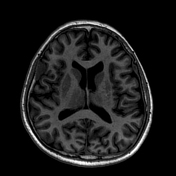

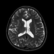

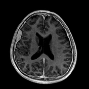
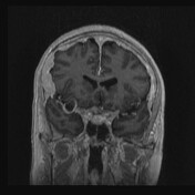
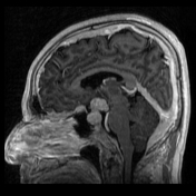

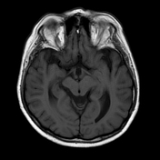
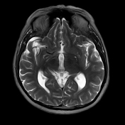
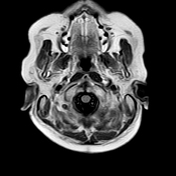
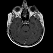
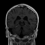
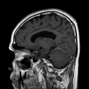
There is widespread dural thickening forming mass-like lesions in the skull base extending through the cisternal spaces, posterior fossa, tentorium cerebelli and subdural space in both brain hemispheres. Another extra-axial lesion occupied the sellar-suprasellar region with infiltration of the pituitary stem and optic chiasm and projection toward the suprasellar cistern. Postcontrast T1WI depicts shows a homogenous vivid enhancement of the lesions and the pachymeninges
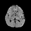
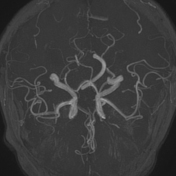
DWI shows no restriction and TOF-MRA shows no occlusions or stenoses of the proximal intracranial circulation.
Histopathological Examination:
Microscopic examination of the specimen revealed a mixed inflammatory infiltrate of mature histiocytes with evidence of emperipolesis, T and B lymphocytes, and plasma cells, showing positive staining for S100 and CD68 and negative staining for CD3 and CD20. The Ki67 index was 15%. The histopathology report revealed Rosai-Dorfman disease.

Brain MRI after 6 months shows almost complete resolutions of the mass-like dural lesions. A focal dural thickening persists at the right tentorium cerebelli and a small enhancing lesion at the pituitary steam at postcontrast T1WI.
Case Discussion
Rosai-Dorfman disease is a rare but well-recognised idiopathic histioproliferative disease affecting the systemic lymph nodes. Isolated intracranial lesions are extremely rare presenting as solitary or multiple extra-axial dural mass-like lesions with vivid homogeneous enhancement.
Differential diagnosis in this case would include the following entities:
- Plaque and malignant meningioma.
- Diffuse pachymeningitis (Tuberculosis, Neurosarcoidosis)
- Meningeal metastasis.
The radiological differentiation from meningiomas is difficult, and can only be achieved with histopathological examination.


 Unable to process the form. Check for errors and try again.