
Presentation
Swelling and weakness of the arm following a fall to the ground
Patient Data


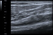
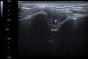
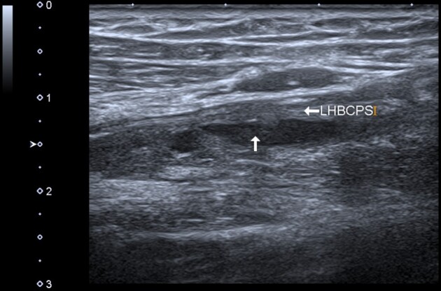
Long head biceps tendon: anechoic fluid occupying regions of tendinous tear and at the myotendinous junction. There is mild subluxation of the long head of biceps tendon within the bicipital groove. A complementary shoulder scan revealed:
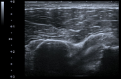
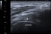
full thickness tear of supraspinatus tendon with anechoic fluid occupying the defect
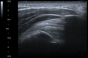
subscapularis tendinosis
osteoarthritic changes of the acromioclavicular joint
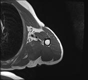
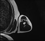
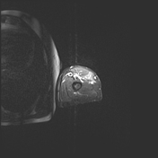
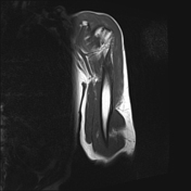
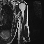
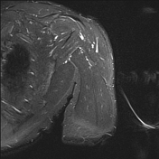
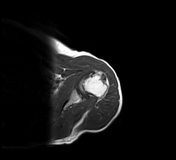
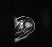
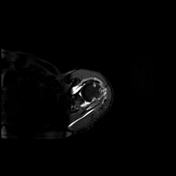
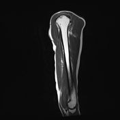
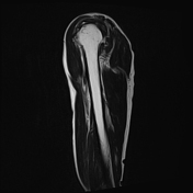
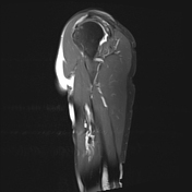
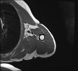
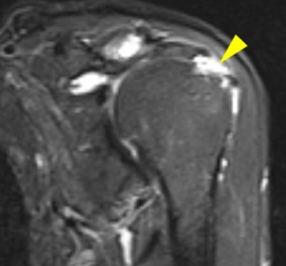
The long head of biceps is replaced by a fluid signal (displaying a high signal in both T2 and STIR images) at the tendon and the myotendinous junction with high grade tear. There is a full-thickness tear of the supraspinatus tendon at its insertion site with high signal in both T2 and STIR sequences. Note presence of subacromion, subcoracoid bursitis and osteoarthritic changes of the acromioclavicular joint.
Annotated images describing the correlation between the findings seen in both ultrasound and MRI:
anechoic fluid within the substance of the long head of biceps (white arrows)
the fluid signal within the tendon (red arrowhead)
the fluid interrupting the distal part of the supraspinatus tendon (labeled)
the high signal in STIR images interrupting the tendon (yellow arrowhead)
Case Discussion
Most cases of long head of biceps tendon tears are secondary to atrophy and rotator cuff muscular pathology. An isolated long head of biceps tendon tear is uncommon. The injury usually involves the upper part of the tendon and at the proximal myotendinous junction. We present a case of a male patient complaining of swelling of the forearm following a fall with the characteristic: "Popeye deformity". Radiological evaluation revealed a ruptured long head of biceps in the form of a near-total tear interruption of the muscle fibers at the tendon and myotendinous junction. Underlying attrition of the rotator cuff was also identified in the form of subscapularis tendinopathy and full-thickness partial-width tear of the supraspinatus.


 Unable to process the form. Check for errors and try again.