
Presentation
Slowly enlarging mass of right thumb for the past 7-8 years, accelerating in growth over the last year. No pain or numbness but affects daily activities due to its size. X-rays and ultrasound were done as patient was unable to tolerate MRI.
Patient Data

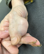
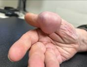
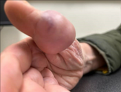
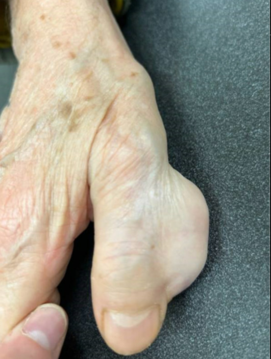
Photographs show a 5.5 x 2.5 x 5.5 cm mass over radial aspect of the right thumb. This was firm, did not transilluminate, and was non-mobile.

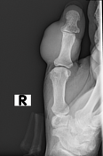
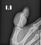
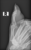
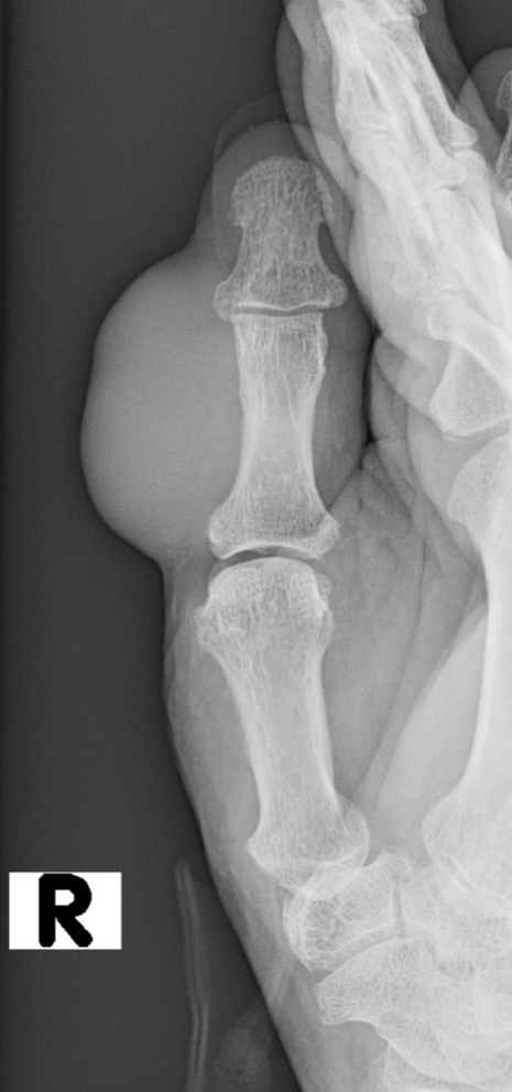
Radiographs show a large soft tissue mass centred around the proximal phalanx of the right thumb without osseous lesion or erosion.
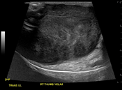
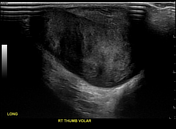
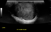
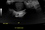
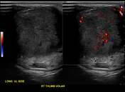
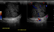

Ultrasound shows a well-circumscribed heterogenous solid mass with peripheral and central internal vascularity on colour Doppler sampling. This is located in the subcutaneous tissues at the radial side of the thumb. There is pseudocystic appearance and acoustic enhancement deep to the mass. There is no obvious association with the flexor tendon. Neurovascular bundle is seen deep to the mass. Adjacent vascularity is confirmed on colour Doppler. No Tinel's sign was elicited.
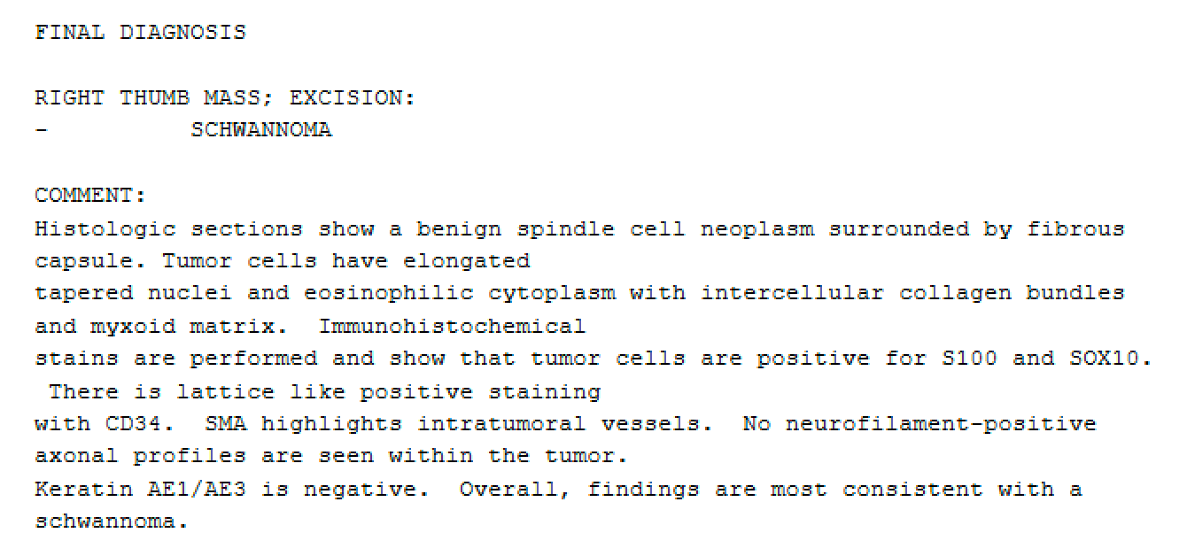
Surgical Pathology report confirming diagnosis of schwannoma
Case Discussion
The mass was surgically resected. The radial neurovascular bundle was associated with the mass and carefully peeled from the mass during surgery. Pathology was consistent with schwannoma. On two-week follow-up, the incision was healing well and there were no impairments in movement or sensation.
Schwannomas have varied sonographic features, although often appearing as encapsulated, hypoechoic masses with characteristics like posterior acoustic enhancement, target sign, pseudocystic appearance, and internal flow 1. However, sonography cannot reliably differentiate between schwannomas, neurofibromas, and malignant peripheral nerve sheath tumours. Peripheral nerve sheath tumours may resemble ganglion cysts due to their hypoechoic features and posterior acoustic enhancement, but internal blood flow observed on Doppler sonography in peripheral nerve sheath tumours can distinguish them 2. Identifying peripheral nerve continuity also suggests the diagnosis of a peripheral nerve sheath tumour but may be difficult to visualise with smaller nerves.
Case co-author: Amirmasoud Negarestani (Loyola University Medical Centre)


 Unable to process the form. Check for errors and try again.