
Presentation
Under evaluation for irregular menses and primary infertility
Patient Data


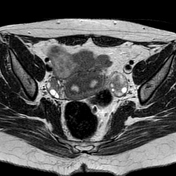
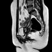
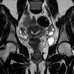
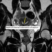
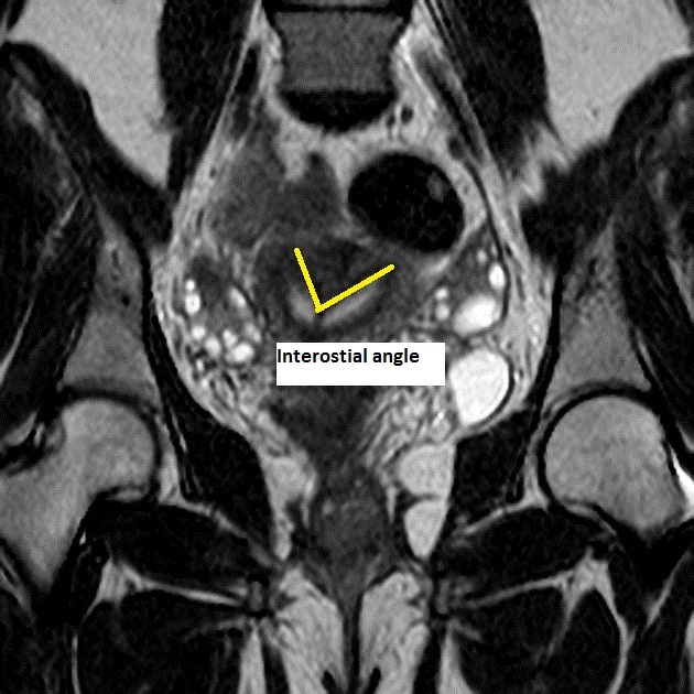
The uterus is normal in size with two uterine cavities separated by a septum of low signal on T2 weighted images with a maintained convex shape of the surface of the fundus with an acute angle between the uterine cavities. There is a common cervical canal with T2 hypointense incomplete vertical septa within the cervical canal.
Depth of internal indentation of septum (from fundus) – 22 mm.
Interostial angle - 60 degrees.
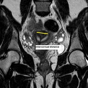
Intercornual distance - 30 mm.

Case Discussion
MRI is the imaging modality of choice for diagnosing uterine anomalies. Septum in this case is composed of fibrous tissue and shows low T2 signal intensity.
The prime differential is the bicornuate uterus, which is differentiated by the shape of the external uterine contour. Bicornuate uterus shows a deep (>1 cm) fundal cleft in the outer uterine contour and an intercornual distance of >4 cm.


 Unable to process the form. Check for errors and try again.