
Presentation
Patient being evaluated for infertility.
Patient Data
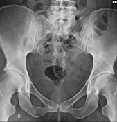
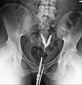
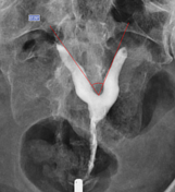
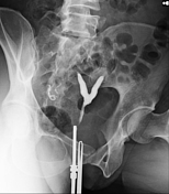
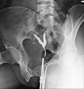
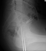
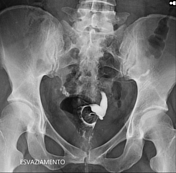
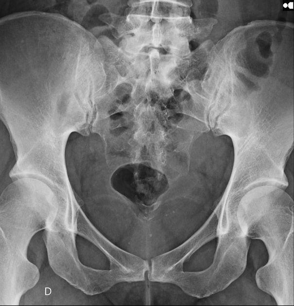
Pelvic radiograph shows radiodense rounded formations in the pelvis, which may be associated with phleboliths. Contrast medium injected through the cervical os progressed satisfactorily to the uterine cavity which exhibits two separate extensions, with narrow intercornuate distance and angle of ~60º (<75º), findings suggestive of septate uterus. Adequate filling of the uterine tubes is observed, which usual morphology but without peritonealization of contrast - representing bilateral tubal obstruction.
Case Discussion
Septate uterus is an anomaly of uterine duplication in which there is failure of fusion of the Müllerian (paramesonephric) ducts, and is the most common congenital uterine anomaly 1. The condition is usually an incidental finding on pelvic examination or found during infertility evaluation 2. The bicornuate uterus may have a similar appearance to the septate uterus, which can cause errors in imaging diagnosis - on hysterosalpingogram, angles > 105º suggest a bicornuate uterus, while angles < 75º suggest a septate uterus 3. Magnetic resonance is the method of choice for diagnosis 1,2. Surgical correction may be considered for reproductive purposes, as it can cause infertility and obstetric complications 1,4.


 Unable to process the form. Check for errors and try again.