
Presentation
Trauma to ring finger (sibling stepped on it)
Patient Data

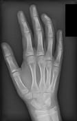
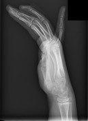
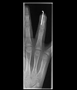
The ring (fourth) finger distal phalanx demonstrates soft tissue swelling and an open fracture in which the physis is separated with apex dorsal angulation and dorsal displacement of the distal fragment. There is a small fracture fragment adjacent to the metaphyseal portion, consistent with a Salter-Harris type II fracture.
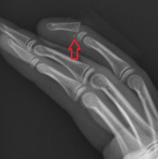
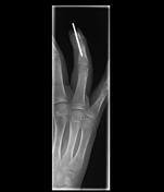
Zoomed in to the ring finger distal phalanx, the lateral radiograph best demonstrates the displaced and angulated physeal fracture and a metaphyseal bony fragment (red arrow), consistent with Salter-Harris type II fracture.
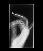
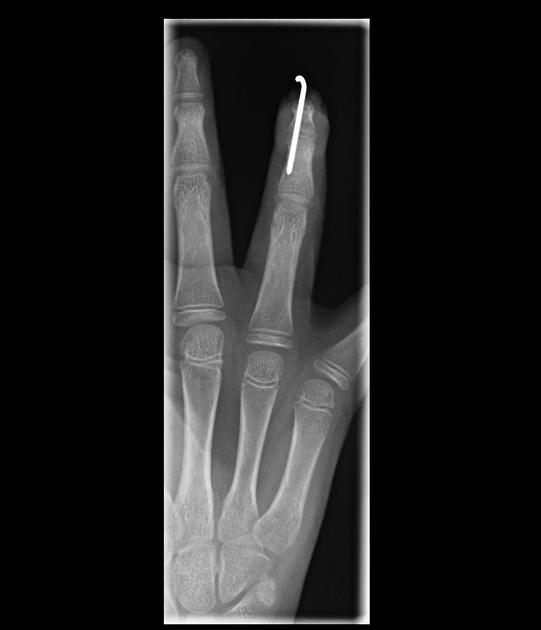
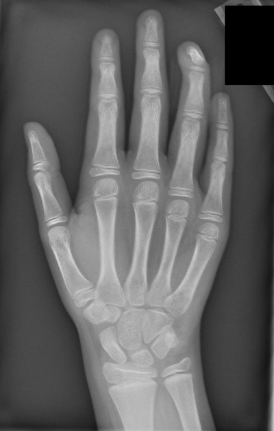
After interval reduction and percutaneous fixation of a Salter-Harris type II fracture of the distal fourth phalanx with a single K wire, the alignment is anatomic.
Case Discussion
At initial presentation, physical examination showed an open injury of the ring finger with flexion deformity of the distal interphalangeal joint and involvement of the nail bed. The clinical and radiographic findings were consistent with a Seymour fracture. Operative management is indicated and the patient immediately underwent irrigation and debridement, open reduction and percutaneous fixation of the fracture. Intraoperatively, the nail plate appeared well adhered to the nail bed, so the nail was left in place and sutured to the proximal skin. Eventually, a new nail grew in underneath and replaced the injured nail without a cosmetic defect.


 Unable to process the form. Check for errors and try again.