
Presentation
Dyspepsia, abdominal pain and rectal bleeding and no history of remarkable weight loss.
Patient Data
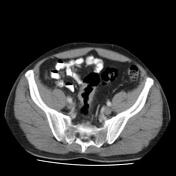

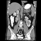
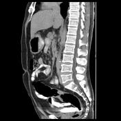
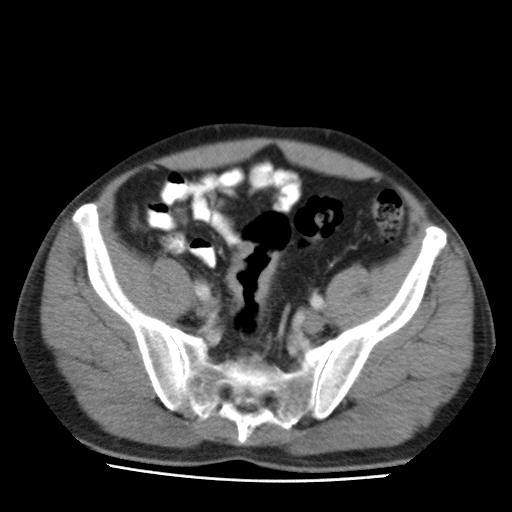
Circumferential mucosal thickening with irregular luminal margin in middle segment of the sigmoid colon is seen. No obvious evidence of pericolic fat extension or significant lymphadenopathy. There is no solid organ metastasis.
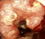
The large ulcerative and fungating mass lesion is circumferentially located in the sigmoid colon.
Endoscopy photo courtesy: Dr Safari Mohamad Taghi (gastroenterologist)
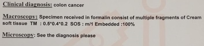

The histology diagnosis of the lesion is adenocarcinoma.
Pathology courtesy Dr Fatemeh Zojajikohan (pathologist)
Case Discussion
The case is a 50-year-old male with a history of dyspepsia, abdominal pain, and rectal bleeding but no remarkable weight loss, the abdomen, and pelvic contrast-enhanced MDCT requested for further evaluation and on the performed MDCT images, long length irregular luminal margin mucosal thickening in sigmoid colon middle segment without obvious pericolic fat extension detected. The patient underwent a colonoscopy and a large ulcerative fungating circumferentially located mass lesion in the sigmoid colon consistent with cancer. The biopsy is done and intramucosal adenocarcinoma is reported.
The sigmoid colon is the most common location of colon cancer and the definitive diagnosis is based on colonoscopy and biopsy but the contrast-enhanced abdomen and pelvic MDCT are routinely requested for the staging of patients with colon cancer. However, MRI is a better option for local staging of the tumor and liver metastasis and dynamic contrast-enhanced MRI and DWI-ADC MRI are promising for further tumor characterization 1,4,5.
Microinvasive surgery, resection of liver and pulmonary metastases, radiotherapy for rectal cancer, and neoadjuvant and palliative chemotherapy all have a limited role in the long-term survival of patients as a result, the colon cancer screening plans are highlighted 2,3.


 Unable to process the form. Check for errors and try again.