
Presentation
Painless shoulder mass noted recently.
Patient Data
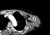

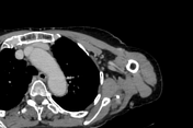
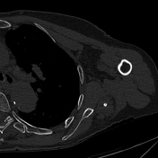
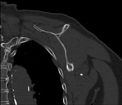
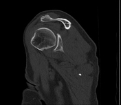

Lobulated soft-tissue mass at the site of patient concern, bounded laterally by the medial border of the long head of triceps brachii muscle, inferiorly by the superior border of the teres major muscle, and superiorly by the inferior border of the teres minor muscle, with the circumflex scapular artery and vein seen at its anterosuperior aspect. The mass measures 45 x 20 x 36 mm (TRV x AP x CC) and is isodense to muscle on the non-contrast scan, with two internal calcified foci. It did not appreciably distort the soft tissue plane, with clear surrounding fat. After contrast administration, it shows progressive moderate enhancement. No prominent feeding artery or draining vein seen, no change in nearby muscle bulk or density, normal nearby bone.
Features of slow flow venous malformation/soft tissue haemangioma of the left medial triangular space.
Case Discussion
Soft tissue haemangiomas are the most common angiomatous lesions and are location-dependent benign vascular soft tissue tumours with a greater female predilection.


 Unable to process the form. Check for errors and try again.