
Presentation
A woman with a 6-year secondary infertility complaint of chronic pelvic pain, and dysmenorrhea. The patient reported a traumatic accident as a child (run over by a car), and subsequent splenectomy.
Patient Data
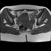

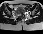
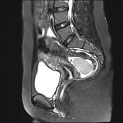
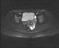
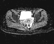

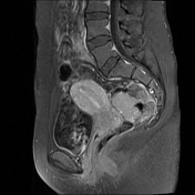

There are multiple enhancing soft tissue nodules of variable size and shapes are seen in the pelvis, two adjacent well‐defined lobulated masses are seen in the pouch of Douglas surrounded by minimal fluid, the larger measures about 5 × 3 cm. Additional few similar enhancing modules are seen scattered throughout the lower abdomen anterior to the bowel.
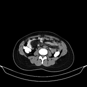
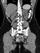
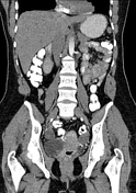
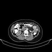

Multiple enhancing variable-sized nodules are seen at the anatomical site of the spleen, the larger seen just lateral to the greater curvature of the stomach, the spleen is missing (history of splenectomy ); other numerous multiple omental and peritoneal enhancing nodules of varying sizes are also seen.
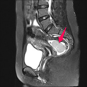
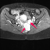
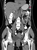
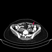
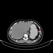

Multiple enhancing nodules (arrows) seen at the anatomical site of the spleen, intraperitoneal, and at the Douglas pouch.
Case Discussion
Splenosis is a benign condition that can occur after splenic trauma or after surgery involving the spleen. These splenic implants are most often seen within the abdominal and pelvic cavities. On imaging, splenosis can be confused with multiple additional entities including metastatic disease, peritoneal carcinomatosis, peritoneal mesothelioma, abdominal lymphoma, or endometriosis depending on its distribution. In all patients with a history of splenic surgery or trauma, splenosis should be on the differential diagnosis of soft tissue nodules in the abdomen and pelvis, especially in the absence of systemic symptoms, to avoid an unnecessary biopsy, chemotherapy, or surgery.
Splenic implants are usually multiple and can be localized anywhere in the peritoneal cavity, but they usually occur on the serosal surfaces of the small and large bowel, the peritoneum, the mesentery, and the diaphragm.
As with accessory spleens, scintigraphy with Tc-99m sulfur colloid or denatured red cells is the most sensitive technique for detecting ectopic splenic peritoneal implants.


 Unable to process the form. Check for errors and try again.