
Presentation
History of a sports injury and pain in the upper left half of the chest.
Patient Data
Age: 15 years
Gender: Male
From the case:
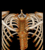
Sternoclavicular joint dislocation


Download
Info
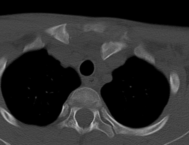
Axial CT showing left sternoclavicular joint incongruence with posterior dislocation of the clavicle.
The dislocated segment of left clavicle is indenting the left brachiocephalic vein.
There is also a medial epiphyseal fracture of left clavicle with bone fragments.
Case Discussion
Posterior sternoclavicular dislocation is a rare traumatic injury that presents a potential risk of injury to mediastinal vascular structures.
The diagnosis is clinical, but x-ray and CT are crucial for better characterisation of the injury.


 Unable to process the form. Check for errors and try again.