
Presentation
New onset of seizures, left-sided port wine stain and facial hypertrophy. Left-sided blindness and abnormal right ophthalmological assessment.
Patient Data

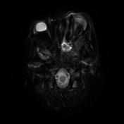
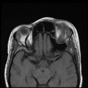
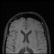
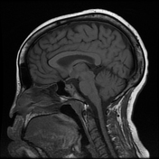
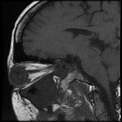
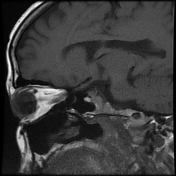
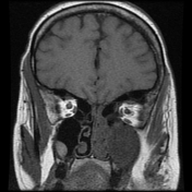
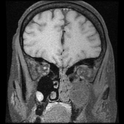
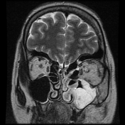
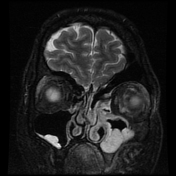

There is left hemifacial, hypertrophy and cutaneous capillary malformation (port wine stain) best visualised on the coronal sequences. There is ipsilateral, chronic, left retinal detachment with a history of multiple previous left orbital interventions and left orbital glaucoma. The left globe is of reduced volume with known blindness. There is a contralateral, right-sided, temporal quadrant, choroidal haemangioma measuring 12.2 x 5.6 mm. The right globe and orbit are otherwise normal.
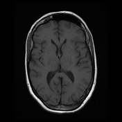
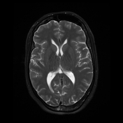
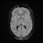
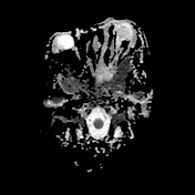
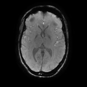
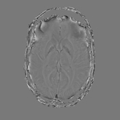
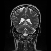
There is left cerebellar hemisphere serpiginous blooming on SWI suggesting cortical and subcortical calcification/ sequelae of regional pial angiomatosis. There is mild regional gliosis and left cerebellar volume loss.
There is incidental chronic pansinusitis with suspicion of fungal sinusitis. There is a heterogeneous appearance to the parotid and submandibular glands bilaterally. There are level 2b and 5a lymph nodes.
Case Discussion
This patient presents with features of Sturge-Weber syndrome with new onset of seizures. She had a background history of unilateral left glaucoma, retinal detachment and multiple ophthalmological surgical interventions which failed to save her vision. She has a left-sided facial port wine stain, and a contralateral right-sided choroidal haemangioma confirmed on dedicated ophthalmologic and MRI assessment.
There are unusual infratentorial, ipsilateral, left cerebellar, cortical and subcortical calcifications. Cerebellar pial angiomatosis has been documented in rare cases of Sturge-Weber syndrome.
In view of the heterogeneous salivary gland appearance, the possibility of fungal sinusitis and the presence of lymph adenopathy, immune suppression and connective tissue disorders were offered as differentials.
Non-contrast CT brain/sinuses and gadolinium administration were contraindicated due to her recent pregnant status (3 months gestation at the time of presentation and imaging).


 Unable to process the form. Check for errors and try again.