
Presentation
Acute onset of headache and dizziness.
Patient Data
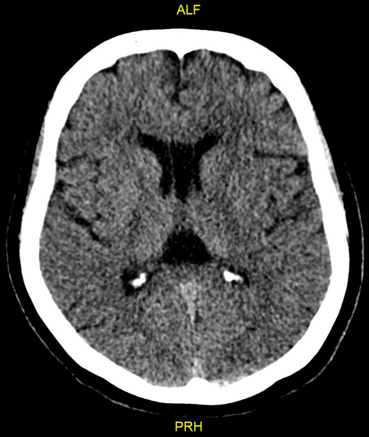
No evidence of subarachnoid haemorrhage on the axial non-contrast CT images.
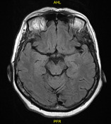

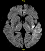
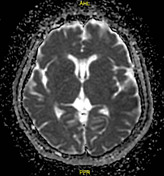
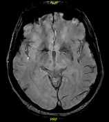
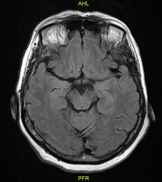
Left parietotemporal areas of recent infarction are seen more appreciated at the insular cortex, associated with ipsilateral multiple linear hyperintensities seen along the subarachnoid space on FLAIR images. These subarachnoid abnormalities show a low signal on SWI images (starting at the bifurcation of the left middle cerebral artery) compared to the contralateral normal side.
Cavum septum pellucidum and cavum vergae are incidental findings.
Case Discussion
The hyperintensities seen along the small vessels in the subarachnoid space on FLAIR are reported in cases of acute ischaemic stroke and are thought to represent slow flow along vessels.


 Unable to process the form. Check for errors and try again.