
Presentation
High blood pressure and headaches.
Patient Data
Age: 60 years
Gender: Male
From the case:
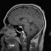
Subependymoma in the fourth ventricle

Download
Info

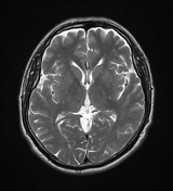
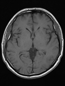
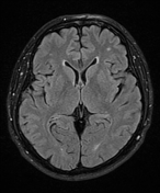
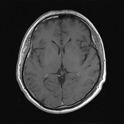
A solid mass is seen in the inferior aspect of the fourth ventricle. Mass shows no enhancement. No hydrocephalus.
Also, there is an incidental arachnoid cyst in the left middle cranial fossa.
Case Discussion
The MRI findings are strongly suggestive of subependymoma of the fourth ventricle.
Subependymomas are uncommon, benign tumours which are usually small, typically less than 2cm in size.


 Unable to process the form. Check for errors and try again.