
Presentation
Post MVA with incidental finding in standard trauma imaging series.
Patient Data
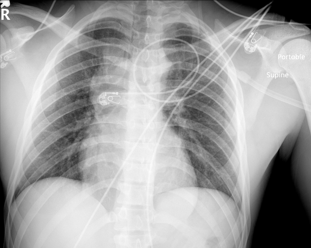
Widening of right superior mediastinal contour.
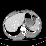

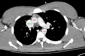
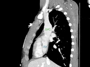
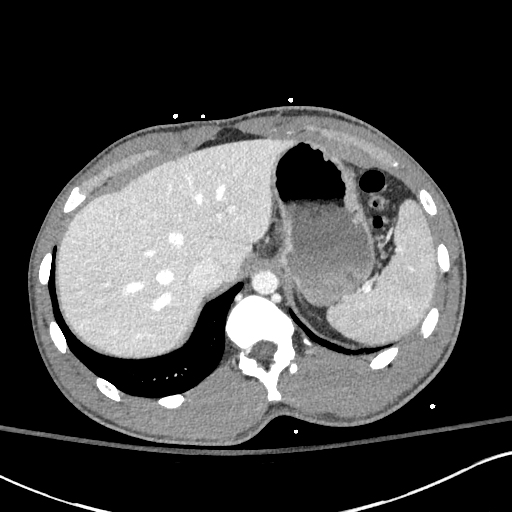
The superior vena cava is wider than the adjacent ascending aorta. No intra-luminal filling defect to suggest thrombosis. No contrast extravasation. No mediastinal fat stranding or hematoma.
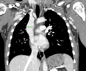
Mild saccular aneurysmal dilatation of the superior vena cava is most obvious in the coronal and sagittal plane.
Otherwise normal. No traumatic injury.
Case Discussion
Radiographic findings of mediastinal widening in trauma settings always warrant further evaluation with contrast-enhanced CT to exclude vascular injury. With advancement in technology and the increased availability of CT imaging more cases of asymptomatic superior vena cava dilatation are being discovered.
Saccular aneurysmal dilatation of the superior vena cava rarely causes complications and typically does not require further management.
This case is submitted with input from:
Jeffery P. Kanne,
MD, FACR, FFCP
Professor (CHS)
Section Chief, Thoracic Imaging
Fellowship Director, Thoracic Imaging.


 Unable to process the form. Check for errors and try again.