
Presentation
Elderly smoker with complaints of right submandibular swelling and dysphagia since 3 months.
Patient Data
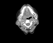

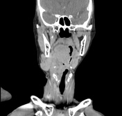
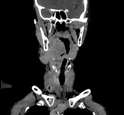

A well defined heterogeneously enhancing lobulated lesion measuring approximately 6.2 x 5.1 x 3.2cm, with few non-enhancing areas of necrosis within, is noted in the right supra-glottic region.
The lesion is involving the right pyriform sinus, the ary-epiglottic fold, the false cord and the right para and retro-laryngeal spaces.
Superiorly the lesion is extending up to the oropharynx at the level of the epiglottis.
Medially the lesion is causing narrowing of the laryngeal airway.
Laterally there is erosion of the hyoid bone and thyroid cartilage in its superior and anterior aspect with exo-laryngeal spread through the thyro-hyoid membrane on right side. No obvious glottic or subglottic extension is noted.
Posteriorly the lesion is seen indenting the proximal oesophagus with resultant collapse of the oesophageal lumen. Inferiorly it is seen to infiltrate the retrolaryngeal space and the trachea-oesophageal groove. The fat plane with the proximal oesophagus is lost. However, the rest of the distal oeseophagus and its lumen appear to be normal.
Laterally the lesion is abutting the right parapharyngeal and posteriorly the retropharyngeal spaces with loss of fat planes with the right submandibular gland. The right submandibular gland appears bulky with focal heterogeneously enhancing areas within suggestive of extension of primary mass.
Multiple enlarged peripherally enhancing necrotic lymph nodes are noted bilaterally at level Ib, II and V largest of size 1.3 x 1.2cm.
The right jugular vein appears dilated.
The nasopharynx is normal.
Bilateral parotid and left submandibular glands are normal.
The paranasal sinuses, visualised orbits and their contents are normal.
Major neck vessels and muscles are normal.
Case Discussion
As the image morphology indicates the mass is extending and invading the tissue beyond the larynx and there are bilateral cevical lymphadenopathy none more than 6 cm in greatest dimension suggests tumour stage-T4a and nodal stage-N2c.


 Unable to process the form. Check for errors and try again.