
Presentation
Long-standing headache with recently increased intensity, which disturbs his daily life.
Patient Data
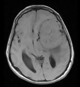

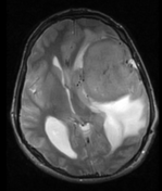
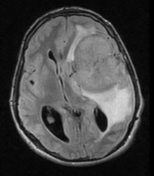
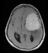
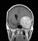
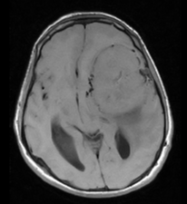
There is a well-defined, extra-axial, altered signal intensity mass lesion, which involves the left frontoparietal and temporal lobes with a positive CSF cleft sign and evidence of displacement of the middle cerebral artery branches and adjacent veins medially. Prominent flow voids within the mass. Returning signals are isointense on T1-weighted, hyperintense on T2-weighted, and FLAIR images (as compared to white matter) with homogenous post-contrast enhancement. Marked surrounding vasogenic edema is seen as well.
Mass effects manifest as compression of the adjacent sulci, left basal ganglia, thalamus, left lateral ventricle, basal cistern, mid-brain, and mid-line shift towards the right (up to 16.0 mm).
The posterior fossa is within normal limits.
Case Discussion
Current MRI features are those of an extra-axial lesion most likely a meningioma. The main differential is that of a solitary fibrous tumor of the dura, although this is far less likely.
The patient has undergone surgery and pathologically confirmed that its meningothelial meningioma.


 Unable to process the form. Check for errors and try again.