
Presentation
Acute left hemiparesis.
Patient Data


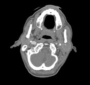
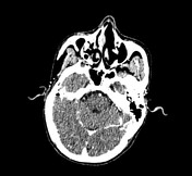
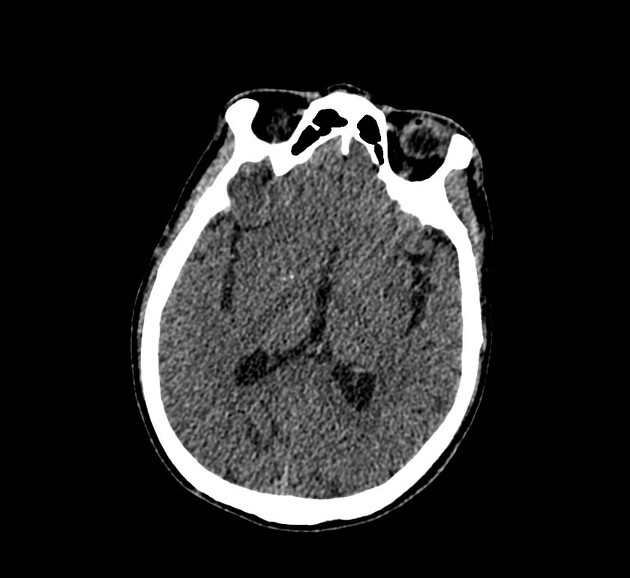
CT non-contrast shows hyperdense right middle cerebral artery in keeping with thrombus. There is a subtle loss of grey-white matter differentiation involving the right insular cortex and the right frontal lobe.
CTA better demonstrates hypoattenuating areas of the right MCA territory and confirms occlusion of the proximal right MCA. There is atherosclerosis and occlusion of the right internal carotid artery origin.
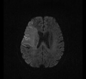
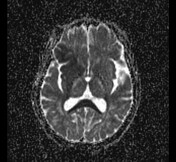
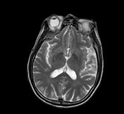
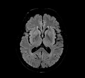
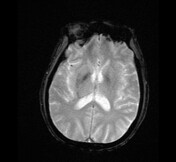
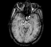
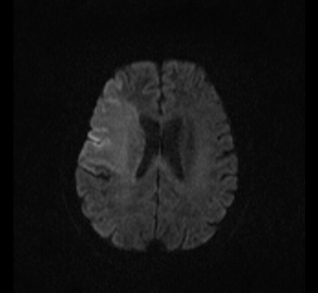
Abnormal hyperintense areas on DWI and subtle high signal on T2/FLAIR involving the right frontal lobe, insular lobe, head of caudate nucleus, posterior limb of internal capsule, are consistent with acute MCA infarct.
Loss of normal flow void of the right ICA and MCA on non-contrast-enhanced MRA.
Note: image quality degraded due to motion artifacts.
Case Discussion
CT and MRI imaging show the simultaneous presence of occlusion ICA and acute MCA thrombosis in keeping with a tandem lesion.


 Unable to process the form. Check for errors and try again.