Presentation
N/A
![](https://prod-images-static.radiopaedia.org/images/59550374/360a1922c3b97bdf42ee9de14e23ec992288c4c528266fb83d1dbd735cc55d70_thumb.jpeg)
![](https://prod-images-static.radiopaedia.org/images/59550375/eeda5e61206bc772163389105d37dd49432390d86ba70d02593ce7cfb992d454_thumb.jpeg)
![](https://prod-images-static.radiopaedia.org/images/59550376/bc003668f82ba04ddf564bedc7e0851d6f775c594075c8c2743df354a8df0462_thumb.jpeg)
![](https://prod-images-static.radiopaedia.org/images/59550377/e644bd5317ab719bb65c5fc94590cceb955b78cb411014d0d5ea71414640e680_thumb.jpeg)
![](https://prod-images-static.radiopaedia.org/images/59558639/977a465337f9da4fde53cc4f2159a5064fc57e751338636dcb1a5746aeacc9fe_thumb.jpeg)
![](https://prod-images-static.radiopaedia.org/images/59550374/360a1922c3b97bdf42ee9de14e23ec992288c4c528266fb83d1dbd735cc55d70_big_gallery.jpeg)
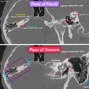
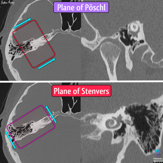
Standard reconstruction planes of temporal bones
![](https://prod-images-static.radiopaedia.org/images/59544053/e5fd2613c4200ba93e2977f482bafc44b7e26bcc6a8930b03e60086bdca50f54_thumb.jpeg)
![](https://prod-images-static.radiopaedia.org/images/59544056/1cebcc8c56bd0aec32cd4158b7ea47fe404eb76ed2545125218bde70065ccde1_thumb.jpeg)
![](https://prod-images-static.radiopaedia.org/images/59550273/61a0ea79834364f4211ee417cd23c9c110c1daf03c0f164a1a1e176f1c9289d3_thumb.jpeg)
![](https://prod-images-static.radiopaedia.org/images/59550274/922a544c9ee518204da3e059321cc6de8dbb57c714eb86b76e1dda2760cc4e1c_thumb.jpeg)
![](https://prod-images-static.radiopaedia.org/images/59544053/e5fd2613c4200ba93e2977f482bafc44b7e26bcc6a8930b03e60086bdca50f54_big_gallery.jpeg)
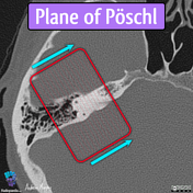
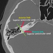
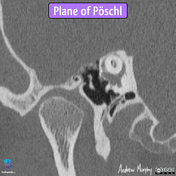
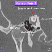
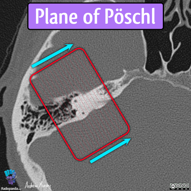
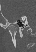
The plane of Poschl is a reconstruction in the same plane as the superior semicircular canal, running roughly perpendicular to the long axis of the temporal bone.
![](https://prod-images-static.radiopaedia.org/images/59550373/4bc239aeaf4d109ce12ff82e7f7fc166d45a4c03e0b0efe57f42cccdbeb433a3_thumb.jpeg)
![](https://prod-images-static.radiopaedia.org/images/59550371/27bd7f194f411e0819e7c098c643ed67e82f77812dbe150737ada17087d620c5_thumb.jpeg)
![](https://prod-images-static.radiopaedia.org/images/59550372/7a163be52e6ac821f78733ecc48ca4ff9af9c5841ffc7334f0c62e85bd406958_thumb.jpeg)
![](https://prod-images-static.radiopaedia.org/images/59550373/4bc239aeaf4d109ce12ff82e7f7fc166d45a4c03e0b0efe57f42cccdbeb433a3_big_gallery.jpeg)
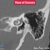
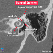
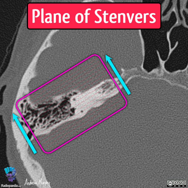
The plane of Stenvers is orthogonal to the plane of Poschl, providing an oblique coronal view that is perpendicular to the superior semicircular canal.
![](https://prod-images-static.radiopaedia.org/images/59550418/39e792a2dbb7bcb41525460a27363856f27d7d5c660fb6a6fdcd8b09c35ef575_thumb.jpeg)
![](https://prod-images-static.radiopaedia.org/images/59550378/24874b201100923aa2a4f5b8d20217bf86ebaa53438ef3c031c6bc70d7304757_thumb.jpeg)
![](https://prod-images-static.radiopaedia.org/images/59550379/71f48114419d1fe6b6a76ae6e9f864c1e0c2caaa872ae4f76eefb735fdb02376_thumb.jpeg)
![](https://prod-images-static.radiopaedia.org/images/59550417/1338ff3229718c6caa74b26a02b7733a07ac857776aaea799f8c0b0b4cbdf7fc_thumb.jpeg)
![](https://prod-images-static.radiopaedia.org/images/59550380/838afa39e6cc5724258919412debc0a22d6f6a1af01301cd6191ed720130d433_thumb.jpeg)
![](https://prod-images-static.radiopaedia.org/images/59550381/283448758adde0241f98fba4567e1d930e8fd0256581217e1c2f548b37312fd7_thumb.jpeg)
![](https://prod-images-static.radiopaedia.org/images/59550418/39e792a2dbb7bcb41525460a27363856f27d7d5c660fb6a6fdcd8b09c35ef575_big_gallery.jpeg)
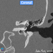
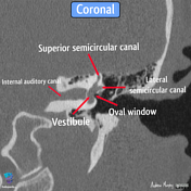
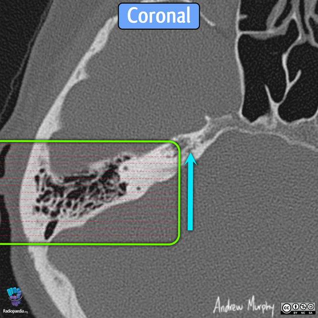
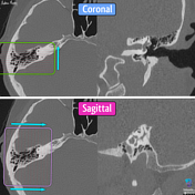
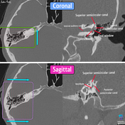
The coronal images should demonstrate the lateral and superior semicircular canals (looking like a crocodile).
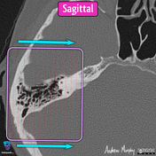
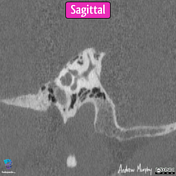
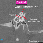
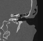
The sagittal is not commonly performed; however, it is orthogonal to the coronal.
![](https://prod-images-static.radiopaedia.org/images/59552633/3e781fbd8a787be60484d8c2a5704fe2ef7ed613c3b742e06b8a7019b774e0bd_thumb.jpeg)
![](https://prod-images-static.radiopaedia.org/images/59552538/662fd6e5f7cd7e6729e2774a605b75506de6b20f1365df5e540877ea77c00e83_thumb.jpeg)
![](https://prod-images-static.radiopaedia.org/images/59552539/179bbfa4ce2377deb3fed6340af2afb902ad2cef5accca41047b9596a5f6ac32_thumb.jpeg)
![](https://prod-images-static.radiopaedia.org/images/59552540/7ac1ea6867377d9ffa982c252b2d0984f8abbc3671df7d776a28e8c6ae1633f7_thumb.jpeg)
![](https://prod-images-static.radiopaedia.org/images/59552541/5027495f29836f2ba951374f57244efda4d7dfaab419870e2340db95a5213544_thumb.jpeg)
![](https://prod-images-static.radiopaedia.org/images/59552542/0e95f96cc8f20b8e8cfff9ebf0693577c524c432954d3df41be910f29b0526a3_thumb.jpeg)
![](https://prod-images-static.radiopaedia.org/images/59552543/a24d28c82b28538d8cd49dd52125f735612dc1e42709d4d9abdf464f2022e0ef_thumb.jpeg)
![](https://prod-images-static.radiopaedia.org/images/59552633/3e781fbd8a787be60484d8c2a5704fe2ef7ed613c3b742e06b8a7019b774e0bd_big_gallery.jpeg)
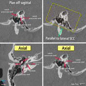
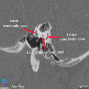
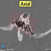
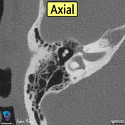
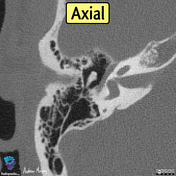
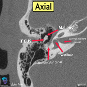
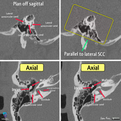
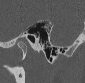
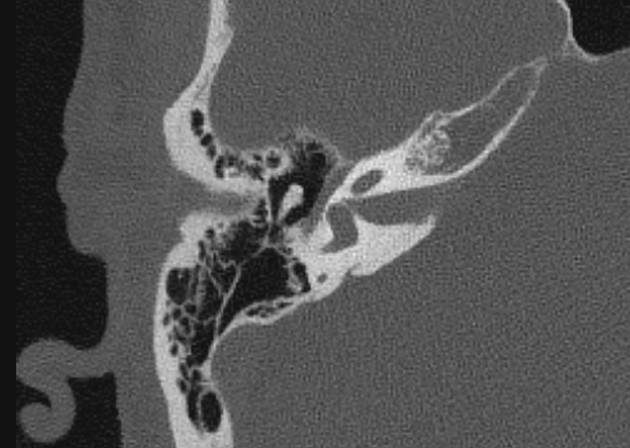
The axial plane is aligned to the lateral semicircular canal (planned off of the sagittal plane).
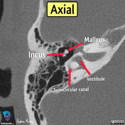
The entire lateral semicircular canal should be visible on a well reformated axial, as well as the middle ear ossicles (the malleus and incus creating the ice cream cone sign).
Annotated anatomy:
![](https://prod-images-static.radiopaedia.org/images/59521753/a56c868dc192c73a25677caf3c3b6e9bd6c826f130434b9d913d89354e3952e0_thumb.jpeg)
![This study is a stack](/packs/stack-YQKLCKBI.gif)
![](https://prod-images-static.radiopaedia.org/images/59523083/a3cff7ec8f0de323a524339aa469651035c00d01f8e285f3f03b200aa3acc372_thumb.jpeg)
![This study is a stack](/packs/stack-YQKLCKBI.gif)
![](https://prod-images-static.radiopaedia.org/images/59522545/c2129744649ac0edba9f9eb6efe22d2dea19c7f2b2ef28d295a7219a83efc9dc_thumb.jpeg)
![This study is a stack](/packs/stack-YQKLCKBI.gif)
![](https://prod-images-static.radiopaedia.org/images/59522886/28ad4a74310a01accd1f4ed5016f92160a2ff086ba2385d24caf55eeef0d3ff1_thumb.jpeg)
![This study is a stack](/packs/stack-YQKLCKBI.gif)
![](https://prod-images-static.radiopaedia.org/images/59522721/bf0b81f6b606f1b9e652bc82887d03d68167e65b803a7aea01e18fc857659c44_thumb.jpeg)
![This study is a stack](/packs/stack-YQKLCKBI.gif)
![](https://prod-images-static.radiopaedia.org/images/59521753/a56c868dc192c73a25677caf3c3b6e9bd6c826f130434b9d913d89354e3952e0_big_gallery.jpeg)
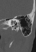
Superior semicircular canal dehiscence best seen on the Stenver plane.
Case Discussion
The plane of Pöschl (in the same plane as the superior semicircular canal) and the plane of Stenvers (perpendicular to the Pöschl plane) are useful reconstructions of the temporal bone in the assessment of the superior semicircular canal. They are different from the coronal and sagittal reformats.
The eponyms can be confusing, so the planes are also referred to as oblique coronals of the temporal bone (which makes sense with the aid of a diagram).
Given the structures are so small, a narrow detector and thin slice size are advisable to image the temporal bone. Care must be taken to reconstruct in these planes for accurate interpretation and reproducibility.