
Presentation
History of recent head trauma, now presented with right facial palsy
Patient Data
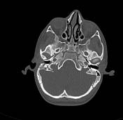

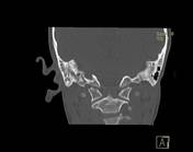

- Branching (V-shaped) fractures are seen traversing the petrous portion of the right temporal bone:
- the longitudinal fracture is seen traversing the mastoid portion of the facial bony canal
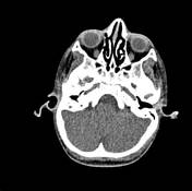
- the transverse fracture is traversing the mastoid air cells and middle ear cavity
- secondary haemotympanum. Intact right ossicular chain.
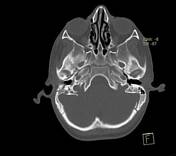
- intact cochlea, vestibule and semicircular canals.
- Fissure fractures are seen involving the lateral aspect of the right occipital condyle and left occipital bone.

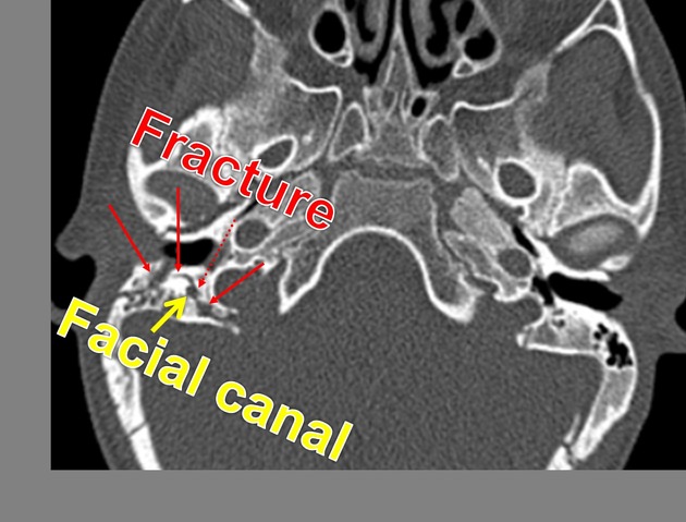
First image: longitudinal fracture (red arrows) is seen traversing the mastoid portion of the facial bony canal (yellow arrow)
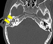
Second image: transverse fracture is traversing the right mastoid air cells and middle ear cavity (yellow chevrons)
Case Discussion
In this case there are mixed temporal bone fractures with longitudinal and transverse fracture components; the longitudinal fracture is traversing the mastoid portion of the facial bony canal which leads to right facial palsy.
Temporal bone fractures are usually a sequela of significant blunt head injury. They can potentially permanently damage hearing and the facial nerve. Early identification of temporal bone trauma is essential to managing the injury and avoiding complications.
Mixed temporal bone fractures are a combination of longitudinal and transverse fracture types, and are probably the most common type. They frequently involve the otic capsule, and are associated with both conductive and sensorineural hearing loss.


 Unable to process the form. Check for errors and try again.