
Presentation
History of fall with trauma to the head and left knee. Complain of headache and dizziness. No loss of consciousness, seizures, or vomiting.
Patient Data


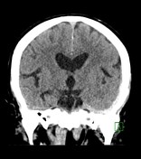
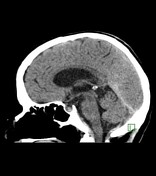
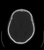
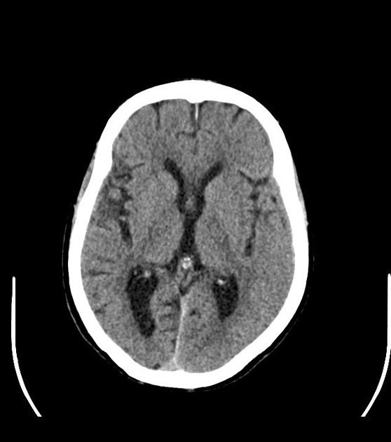
A hyperdense extra-axial collection measuring 7 mm in thickness, keeping with acute subdural haematoma, is seen along the left tentorium cerebelli. Minimal extension of this haematoma under the left squamous temporal bone is noted. No skull fracture, haemorrhagic cerebral contusions, subarachnoid/intraventricular haemorrhage, mass effect, or midline shift is noted. Some hyperdense contents are seen along the anterior interhemispheric fissure which is likely calcifications. Mild age-related atrophic brain parenchymal changes. Right maxillary sinus mucous retention cyst and non-pneumatised right frontal sinus; the remaining paranasal sinuses as well as bilateral mastoid air cells are well-aerated and clear.
Case Discussion
The diagnosis of tentorium cerebelli subdural haematoma can be challenging. It is subtle on the axial plane due to the oblique orientation of the tentorium in comparison to the imaging plane; however, it can be easily diagnosed on coronal and sagittal planes. Therefore, in trauma patients, multiplanar imaging of the brain and review of all available imaging planes play a crucial role in its diagnosis.
The patient was admitted for observation under the neurosurgery services and was later on discharged in a stable condition with GCS 15/15 after 24 hours.


 Unable to process the form. Check for errors and try again.