
Presentation
The patient complains of left orbital pain and squint.
Patient Data


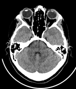
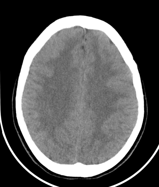
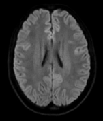
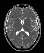
normal size and configuration of the ventricular system
no midline shift
no definite intra-axial areas of abnormal density
unremarkable posterior fossa structure
no definite orbital lesions could be seen on both sides
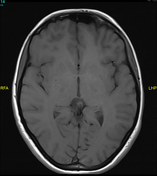
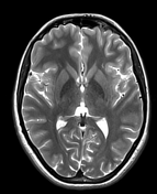
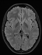
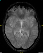
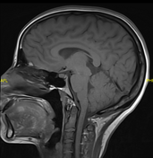
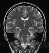
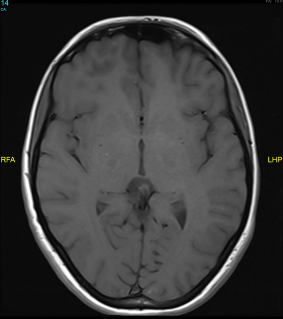
The suspected area of abnormal density on CT at the left cavernous sinus and extends to the left orbital apex is more obvious on MRI and reveals isointense T1 signal, slight hypointense signal at T2 and FLAIR and causes mild attenuation of the cavernous left ICA and increases the distance between the lateral wall of the left cavernous sinus and ICA. Mild periarterial high T2/FLAIR signal is seen also on the left side.
Otherwise, unremarkable MRI brain and orbits.
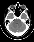
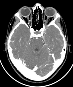
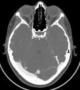

CT with IV contrast confirms the filling defect in the left cavernous sinus which extends to the left orbital apex and revealed no appreciable enhancement could be seen.
Opacified with normal calibre superior ophthalmic vein (no thrombosis).
No carotid-cavernous fistula.
Case Discussion
The clinical data (ophthalmoplegia and squint, which is mostly due to abducent nerve involvement) and radiological findings suggest Tolosa-Hunt syndrome. After corticosteroid therapy, there was a dramatic improvement in the patient's symptoms.


 Unable to process the form. Check for errors and try again.