
Presentation
Vomiting, headache, and seizures. History of previous attacks of meningitis.
Patient Data
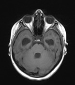

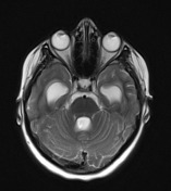
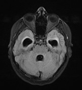
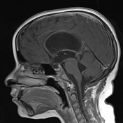

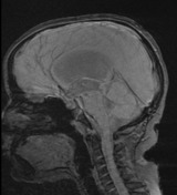

MRI demonstrates marked dilatation of the supratentorial ventricular system noted with associated enlarged 4th ventricle, associated with a thin web at the distal portion of the aqueduct, causing marked tight distal stenosis and with proximal funnelling. This is associated with decreased mamillopontine distance (3 mm) and periventricular T2/FLAIR hyperintensity reflecting associated transependymal permeation.
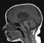
A thin web is also noted at the 4th ventricular outlet foramina, notably the median foramen of Magendie, best appreciated on sagittal thin cuts heavy T2 (CISS) with consequent supratentorial ventricular dilatation and mildly dilated 4th ventricle.
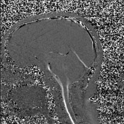
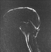
Dynamic CSF flow study and ultra-thin 3D T2 Sagittal sequences revealed: very poor/absent CSF flow signal across the aqueduct and 4th ventricle.
Case Discussion
Multiple webs noted at the distal portion of the aqueduct and 4th ventricle outlet foramina, are suggestive of post-meningitic sequelae. Subsequent active obstructive tetraventricular hydrocephalic changes are noted with transependymal permeation. The absence of flow-void signal intensity on sagittal T2 images at the aqueductal level is a suggestive sign of aqueductal stenosis. MRI CSF flow study shows absent flow through the dilated 4th ventricle in keeping with trapped 4th ventricle.


 Unable to process the form. Check for errors and try again.