
Presentation
Past history of head trauma, presented with painless pulsatile swelling of the left parietal region.
Patient Data
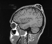

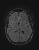
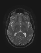
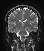
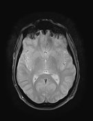
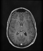
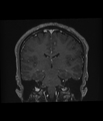
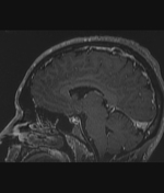
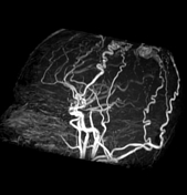
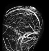

The MRI sequences demonstrate dilated tortuous flow void vessels beneath the scalp of the left parietal region, representing an arteriovenous malformation (AVM). Note dilatation of the superficial temporal artery, and its parietal branch as well as the occipital artery supplying the nidus of the AVM. The draining veins are also visualized. No underlying bony abnormality nor communication with the intracranial circulation.
Case Discussion
MRI features of a traumatic scalp arteriovenous fistula/malformation.
The majority of the vascular lesions involving head and neck soft tissue are congenital, rarely of traumatic origin 1. Usually supplied by the superficial temporal, and occipital arteries (as in this case), and drained through scalp veins or via dural sinuses 1.
CT angiography is very useful in such cases. MRI aids to detect if there is any intracranial extension 1. The treatment is surgical (complete excision) 1.


 Unable to process the form. Check for errors and try again.