
Presentation
A palpable mass on the anterior surface of the left wrist, tender on palpation. History of a left wrist injury that was sutured one year ago. Ultrasound shows a mass continuous with the median nerve. Electromyography (EMG) results indicate severe damage to the median nerve.
Patient Data


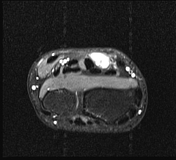
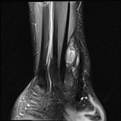
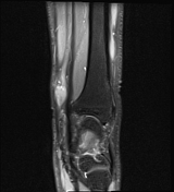

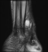


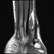
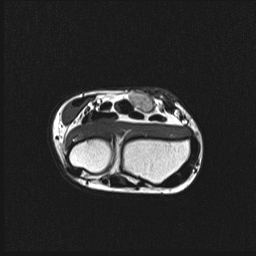
On the MRI, a subcutaneous mass was noted on the anterior aspect of the left wrist, measuring approximately 11 x 6 x 13 mm, with well-defined margins, a fusiform shape, and continuity with the median nerve. The lesion exhibited low signal intensity on T1-weighted images, heterogeneous high signal intensity on T2-weighted images and PDFS, and restricted diffusion.
Surgical report:
a previous scar was noted on the anterior aspect of the lower third of the left forearm, measuring approximately 5 cm, which was well-healed with no signs of dehiscence. On palpation, a mass was felt beneath the scar, approximately 2 x 2 cm in size, with well-defined borders
an incision was made on the lower third of the left forearm, and the tissue was dissected layer by layer to expose the median nerve neuroma, which was adherent to the skin on the anterior forearm; the median nerve was found to be discontinuous at the periphery
a segment of the saphenous vein, approximately 20 cm in length, was harvested. The vein was longitudinally incised and wrapped around the neuroma
Case Discussion
The imaging findings, clinical presentation, and electromyography results are consistent with a traumatic neuroma of the median nerve. The patient subsequently underwent surgical excision of the median nerve and wrapping of the neuroma with a saphenous vein graft. Post-surgery, the clinical symptoms improved.


 Unable to process the form. Check for errors and try again.